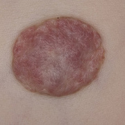
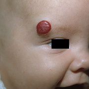
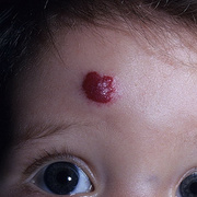
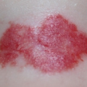
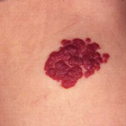
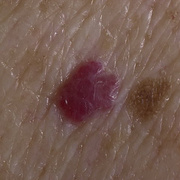
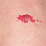
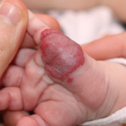
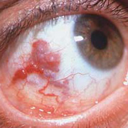
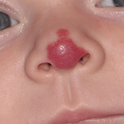
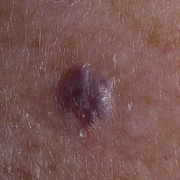
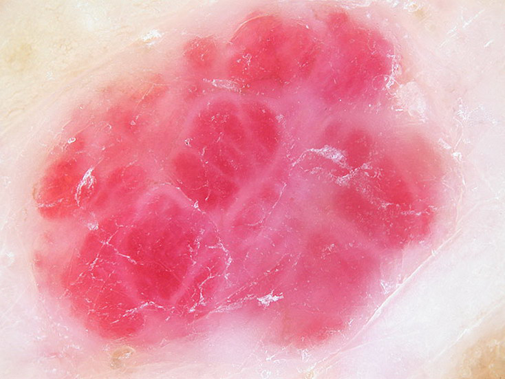
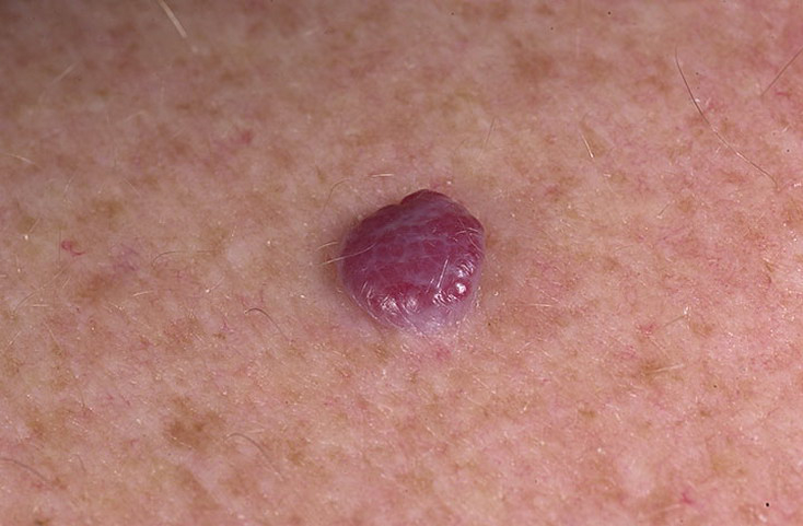
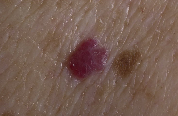
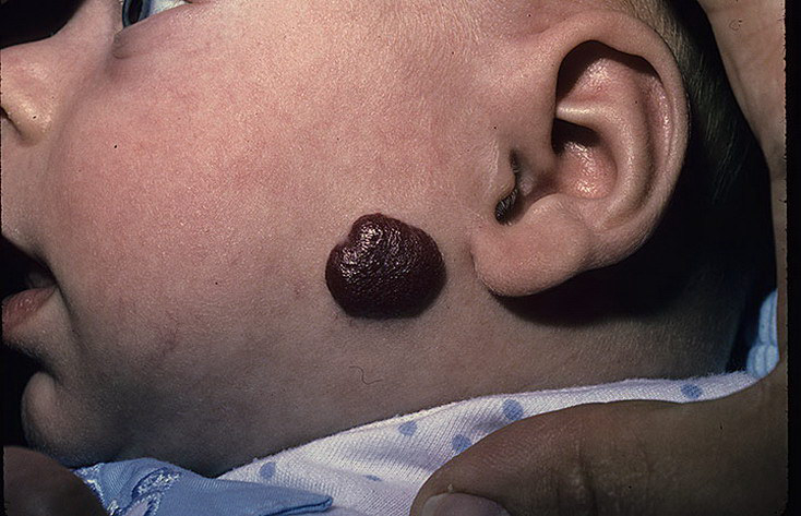
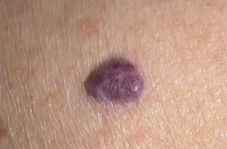
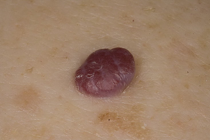
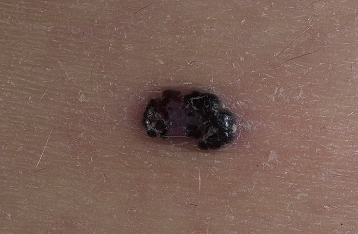
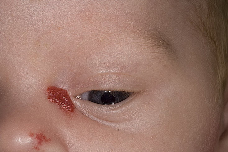
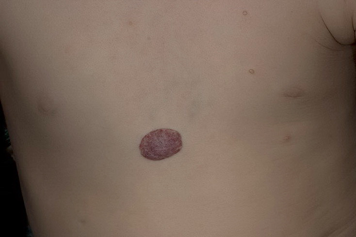
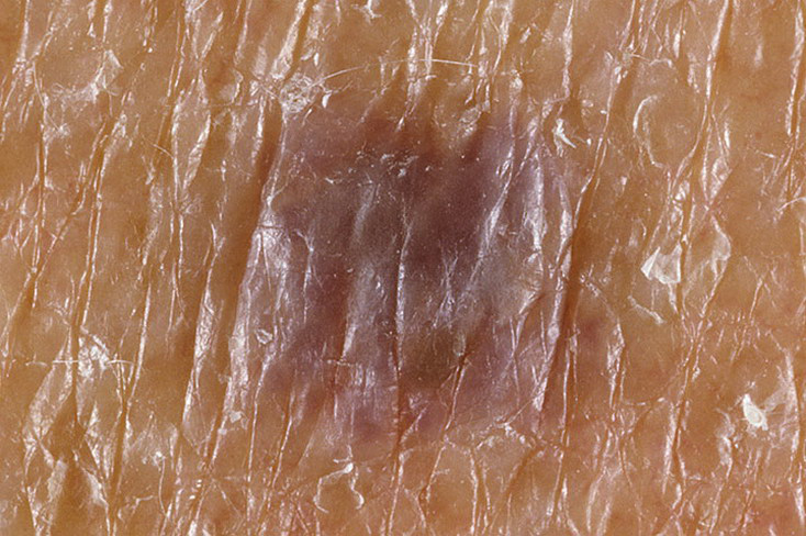
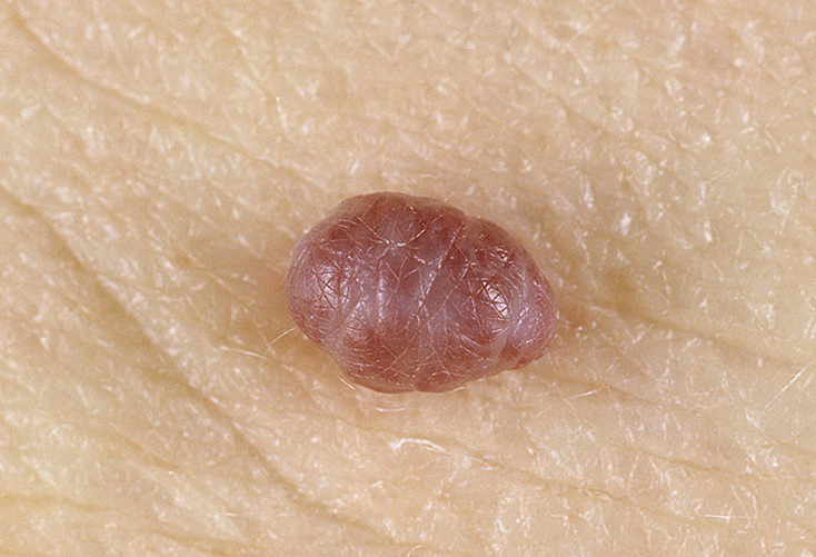
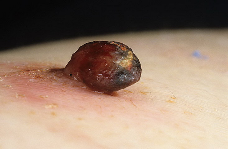
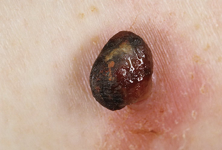
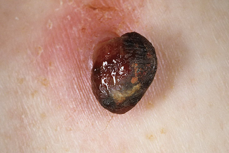
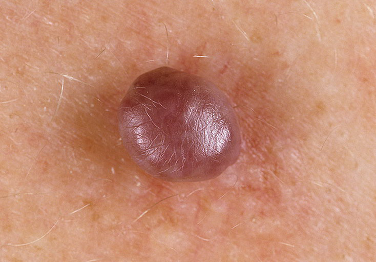
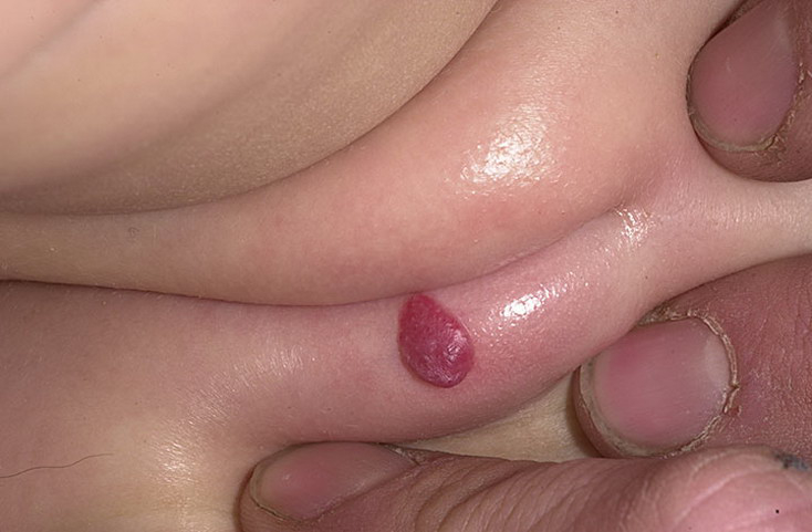
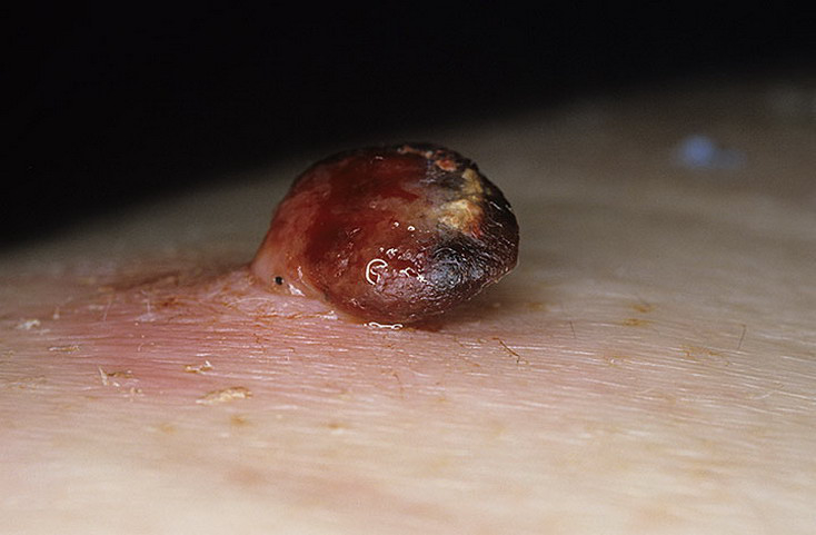
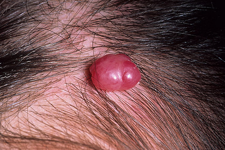
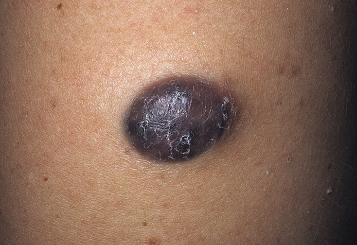
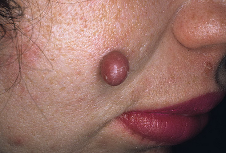
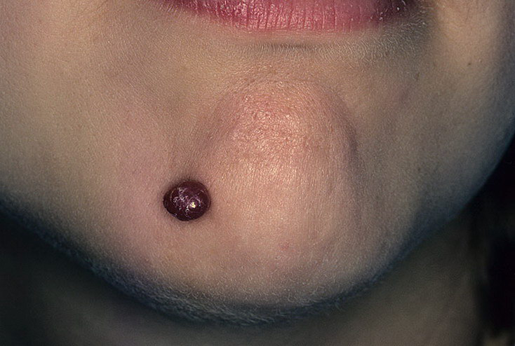
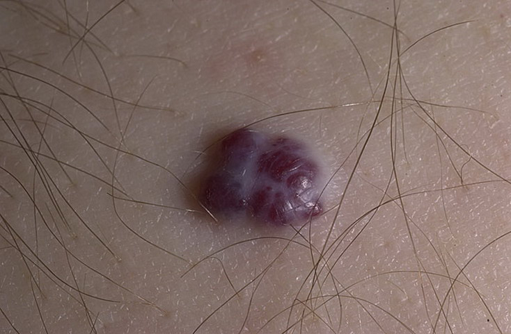
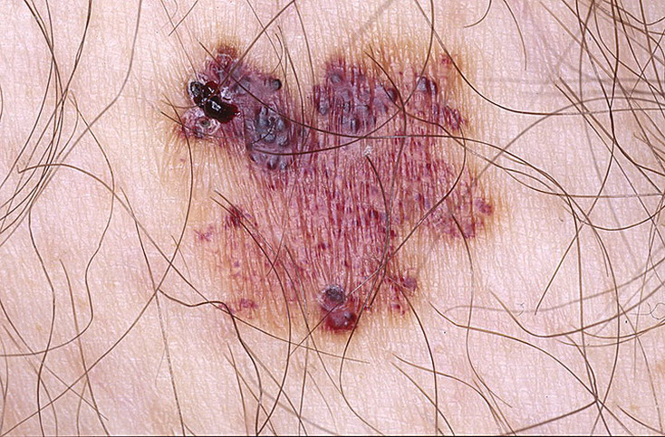
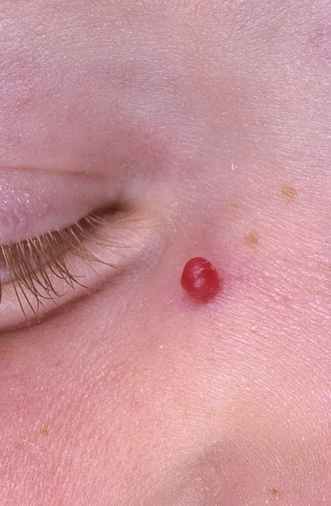
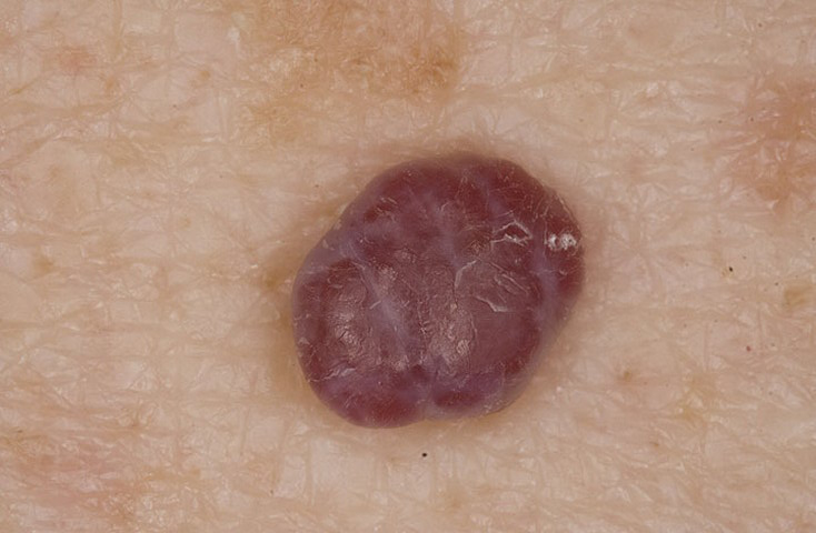
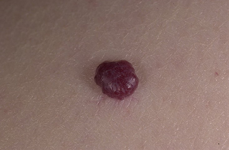
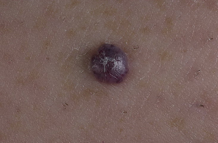
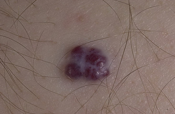
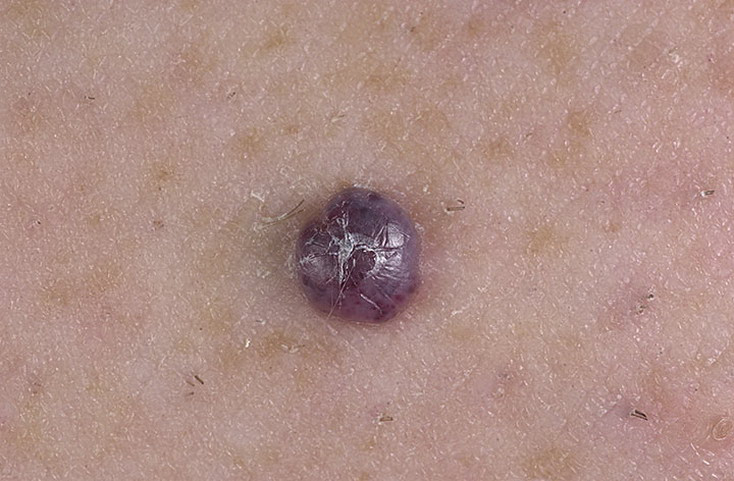
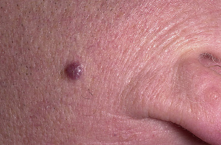
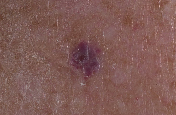
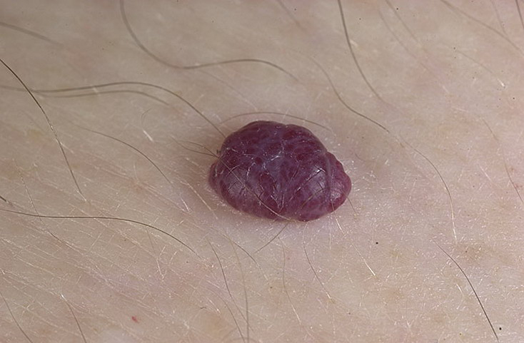
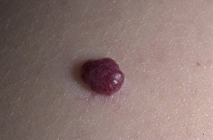
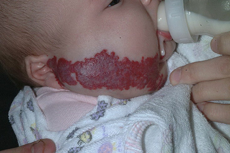
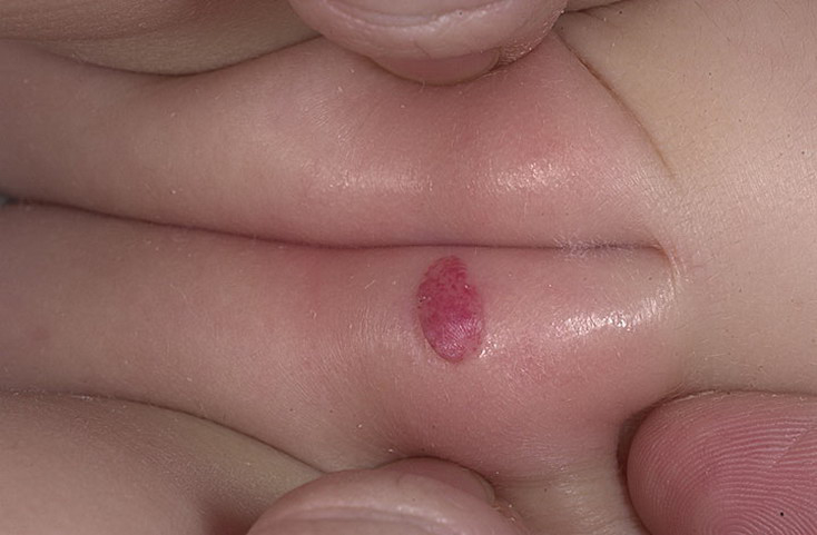
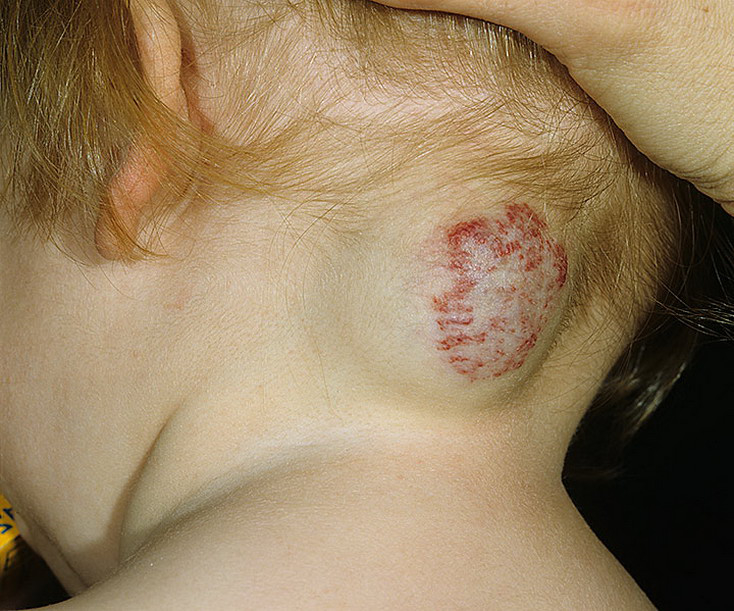
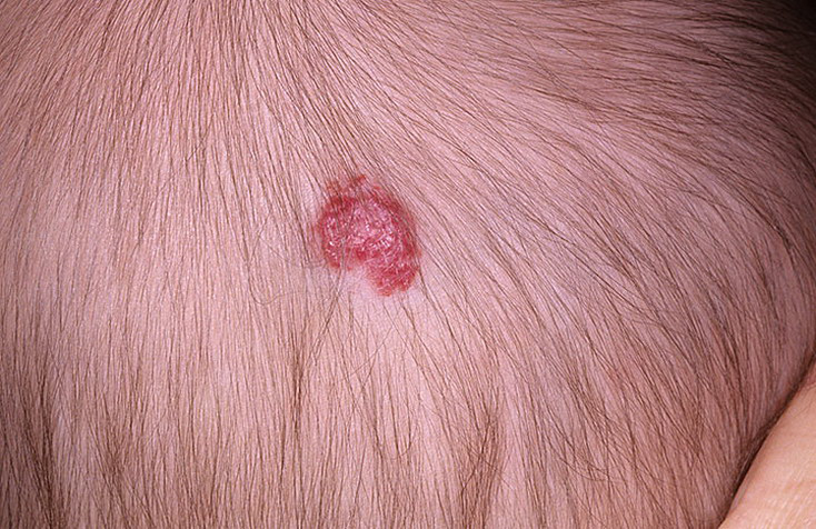
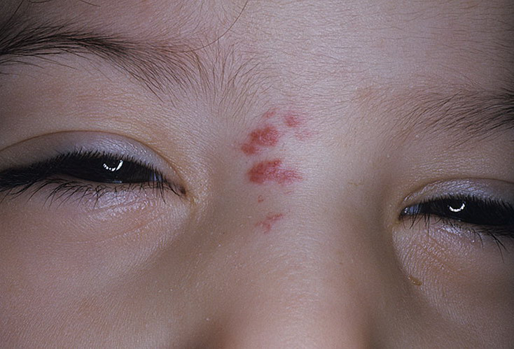
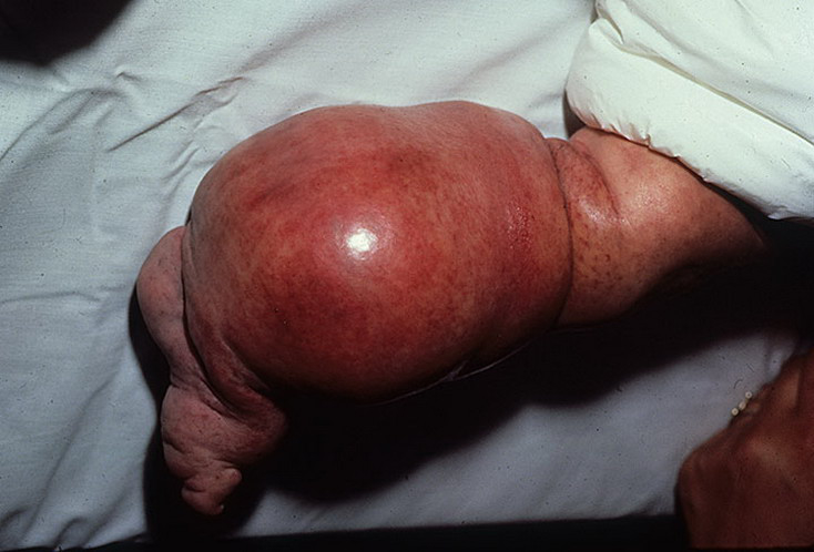
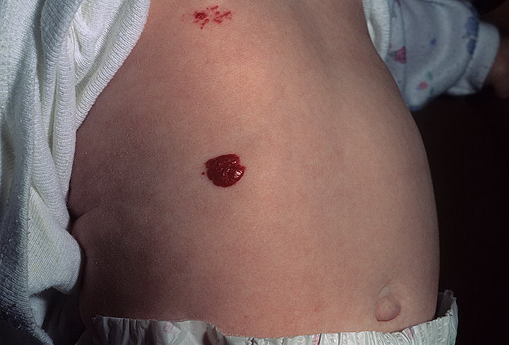
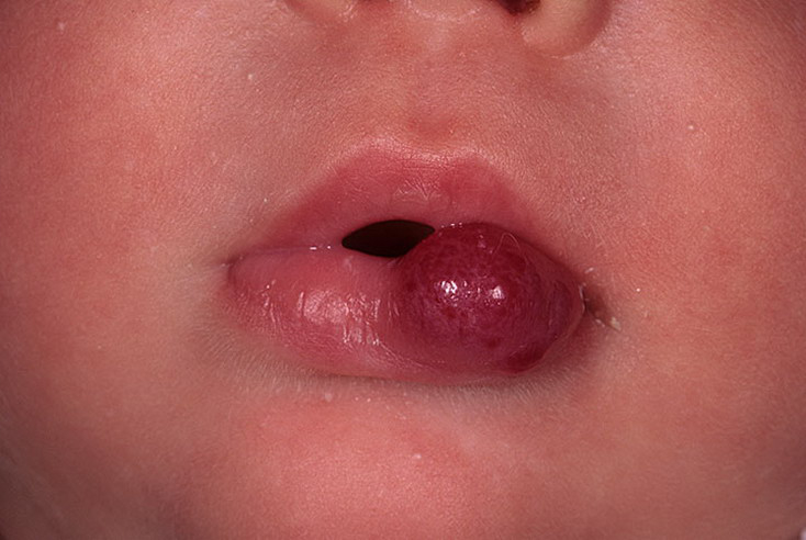
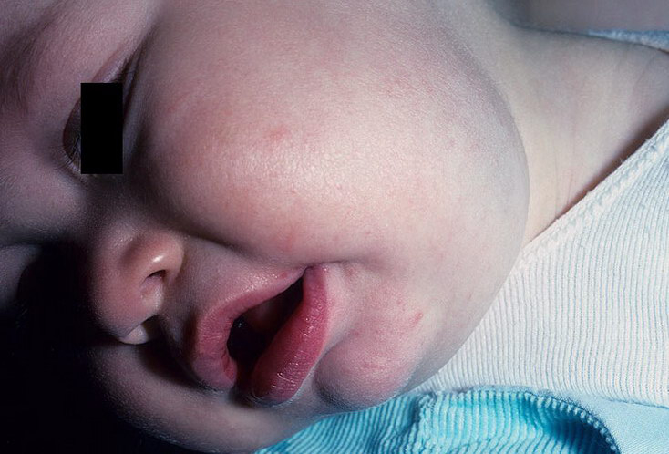
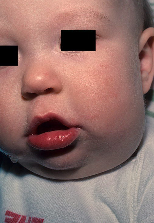
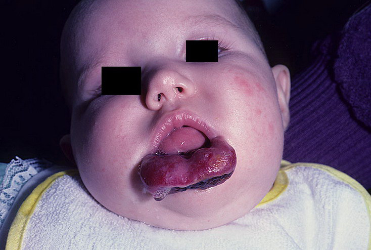
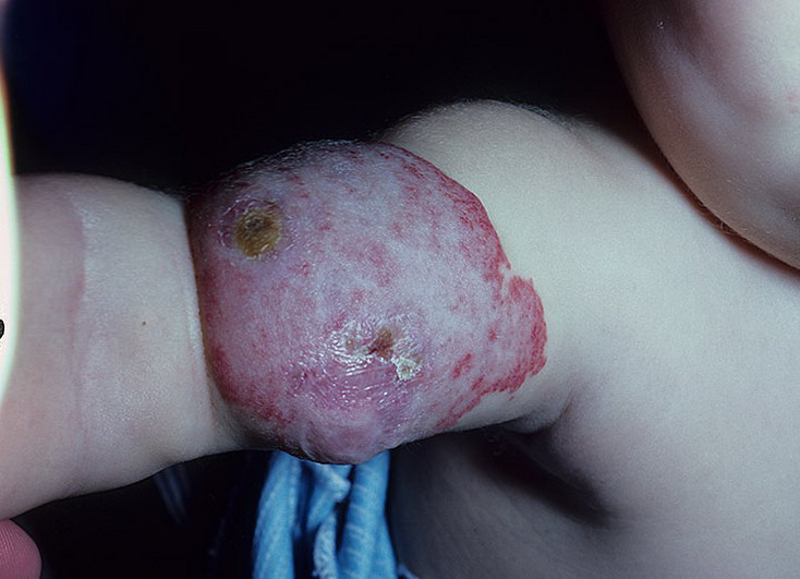
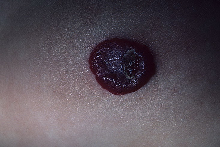
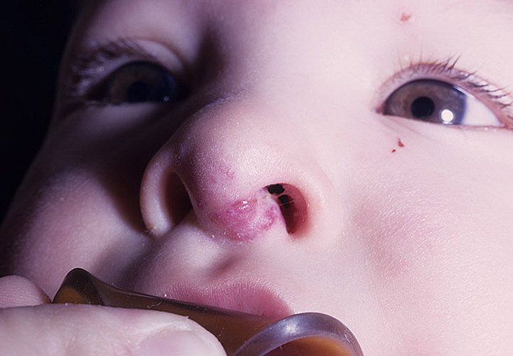
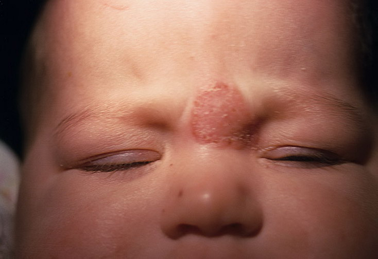
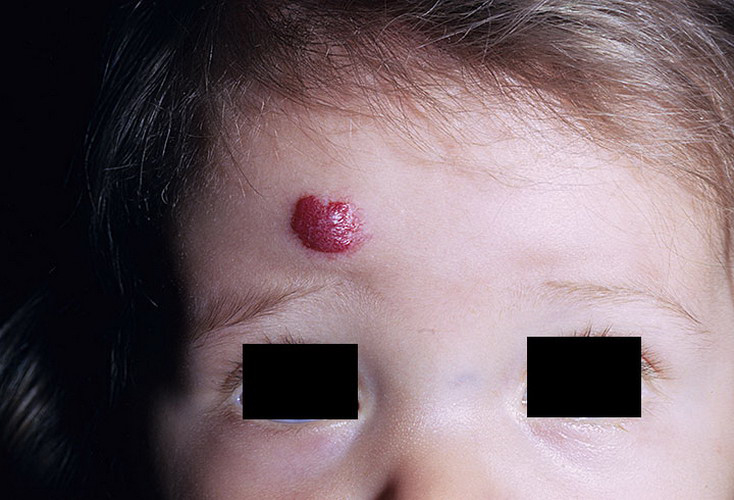
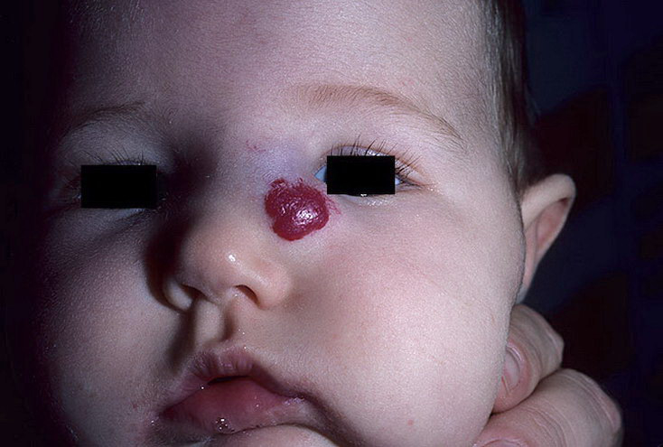
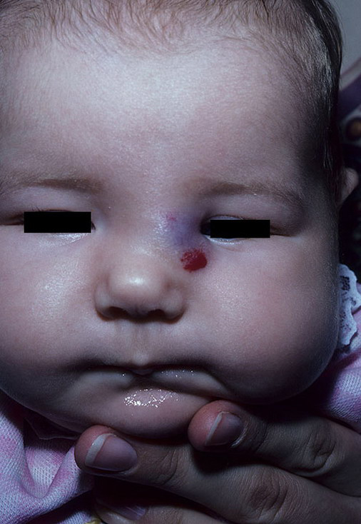
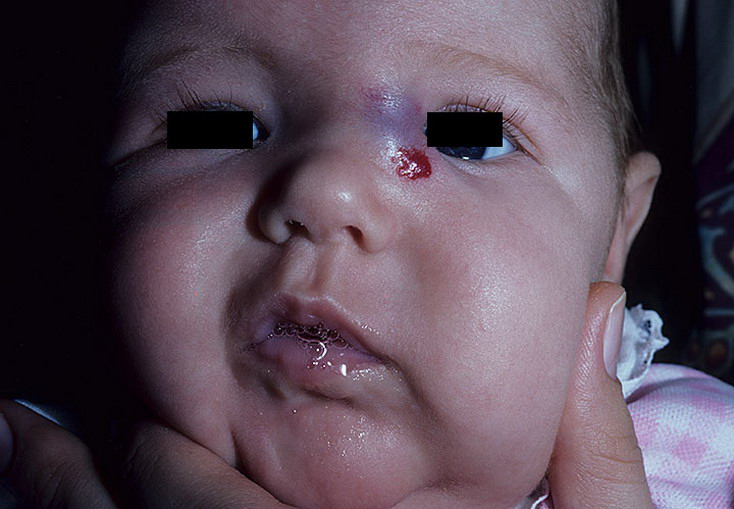
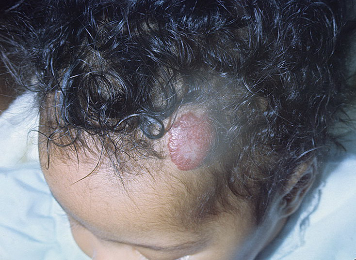
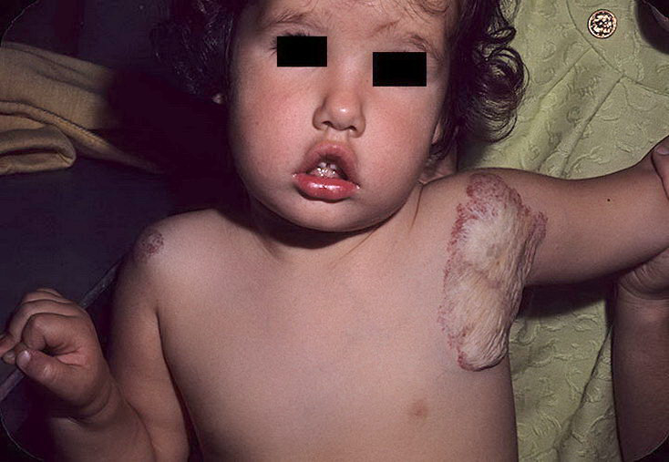
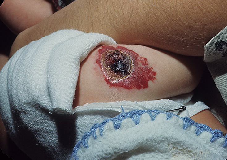
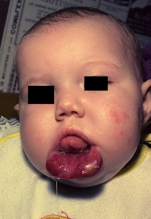
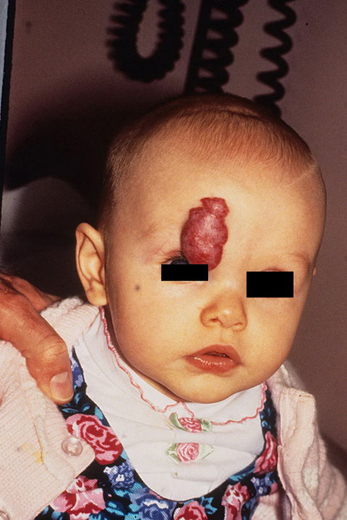
Vascular Hemangioma Pictures - 118 Photos & Images


Also known as “Cavernomas” and “Cavernous Malformations”, These lesions have been found with increasing frequency over the past few years because of the incredible accuracy of MRI scanning. They arise from the tiny vessels that separate the arterial system from the venous system of the brain. There is some controversy as to whether the cavernomas are true vascular malformations or very slow growing tumors of capillary blood vessels. They are characterized as truly benign lesions, and can be completely removed and cured through surgery. However, as many of these lesions are found coincidentally, they are often left alone if no evidence of hemorrhage is present at the time of the MRI study.
They may induce seizures; occasionally, their removal leads to seizure control when medical therapy fails. When they are noted along with hemorrhage, they most often do not cause neurologic devastation, as do brain aneurysms and AVMs. The reason for this has to do with the very low vascular pressure within these malformations.
Consequently, the usual volume of hemorrhage is small, causing temporary deficits that generally improve, although not completely. We now know that the cavernomas do grow slowly over time, and that once they hemorrhage, they tend to do so again. Each subsequent hemorrhage is usually followed by a stepwise deterioration in neurologic function, causing the patient to lose something with each bleed. When they are found in the brainstem, the most compact and important part of the brain, cavernomas may represent a threat to a patient’s life. As might be expected, even a 5cc hemorrhage into the jam-packed area of the brainstem may cause difficulty swallowing, double vision, loss of facial function, and even loss of consciousness.