Hemangioma in Adults Pictures - 62 Photos & Images


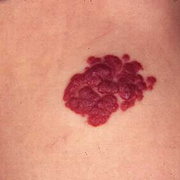
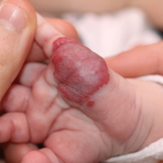
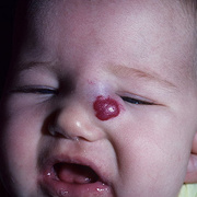
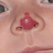
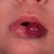
A hemangioma is an abnormal proliferation of blood vessels that may occur in any vascularized tissue. Considerable debate exists as to whether these lesions are neoplasms, hamartomas, or vascular malformations. Mulliken strongly supports classification of hemangiomas as neoplasms, whereas Godanich and Capanacci seem to favor a hamartomatous classification.[1, 2] There seems to be consensus that the term "hemangioma" should refer to "hemangiomas of infancy," which have a predictable natural history that includes absence at birth followed by a period of growth over 6-18 months and then a period of involution that may take several years. "Hemangiomas" affecting the musculoskeletal system are more accurately termed "vascular malformations." These are present from birth and do not involute spontaneously.
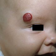
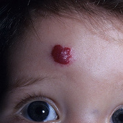
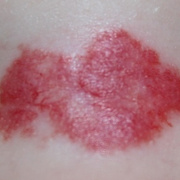
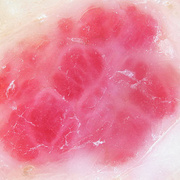
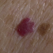
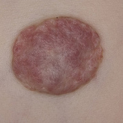
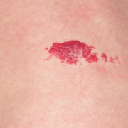
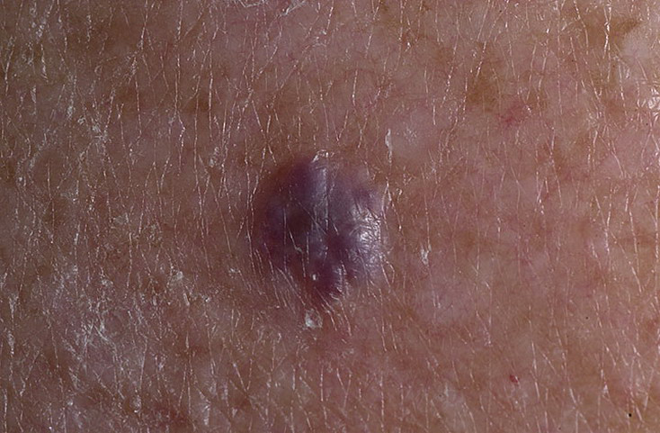
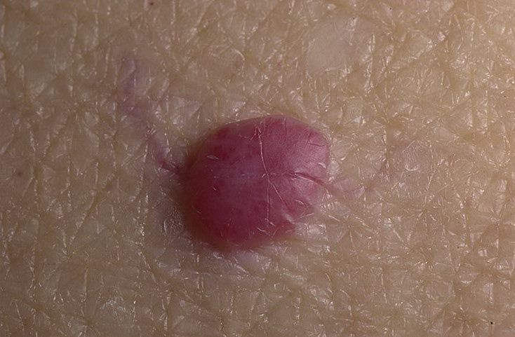
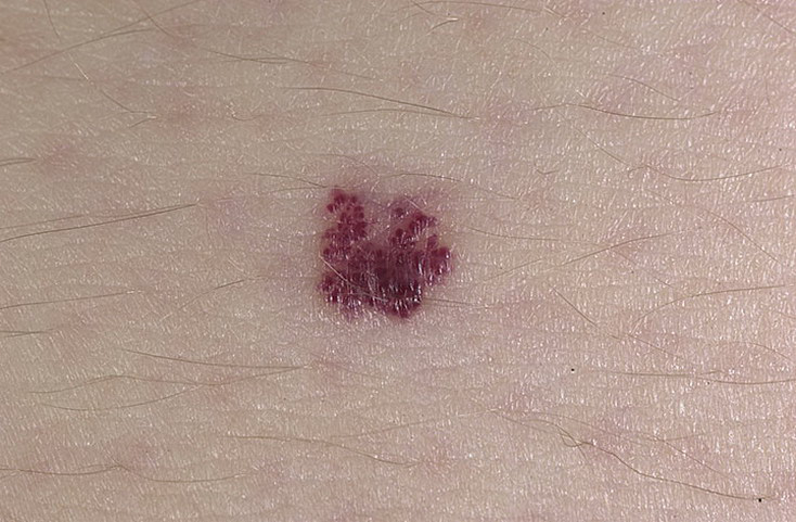
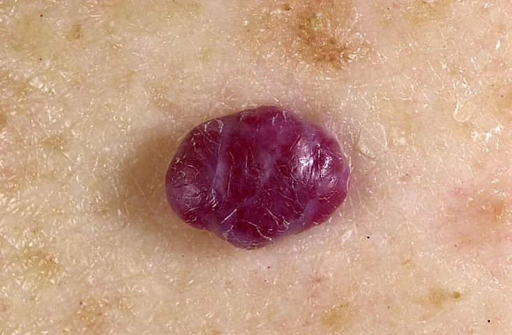
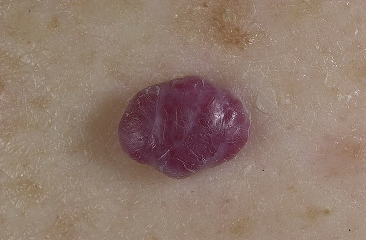
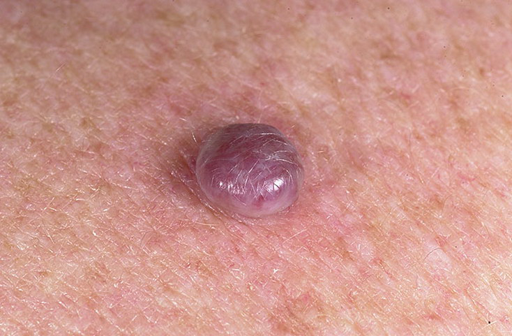
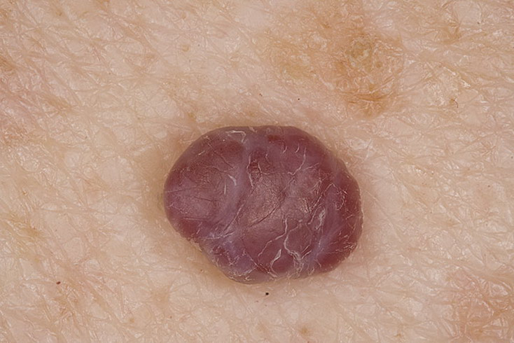
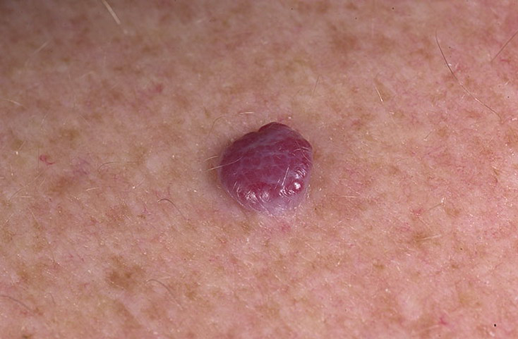
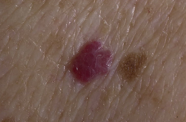
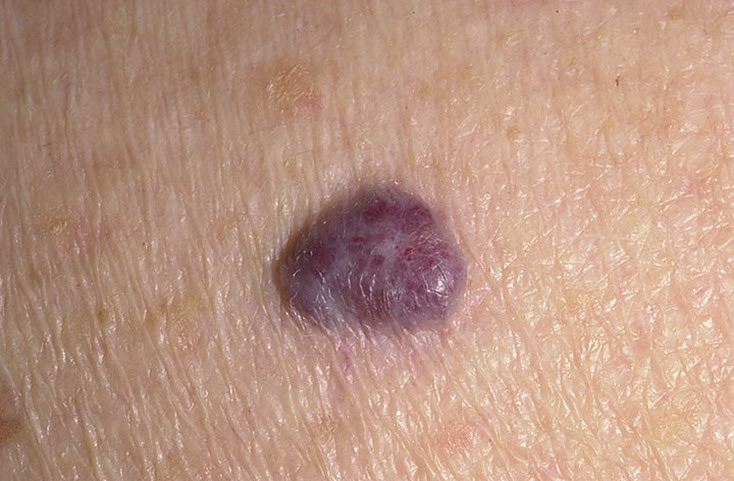
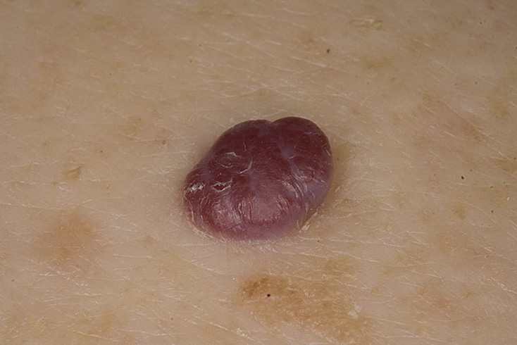
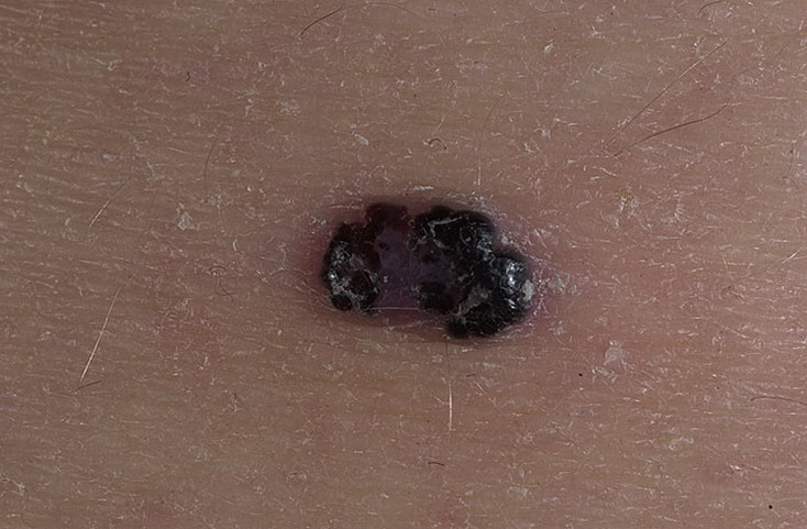
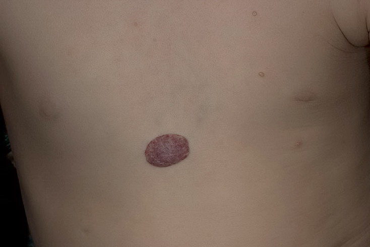
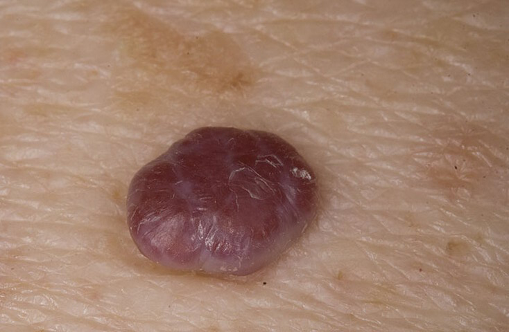
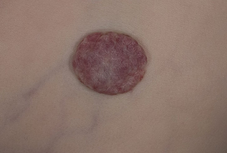
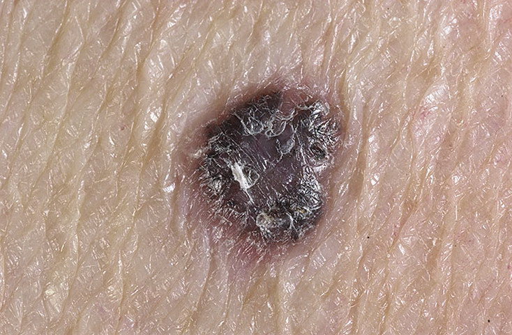
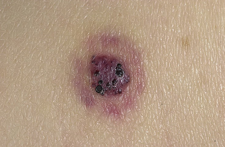
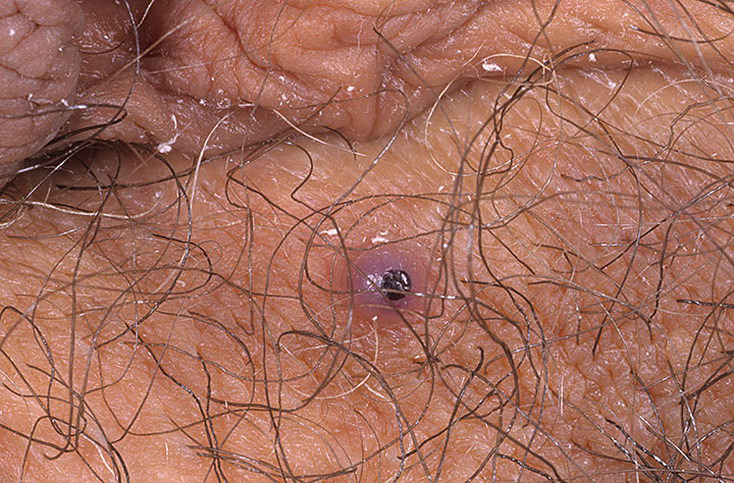
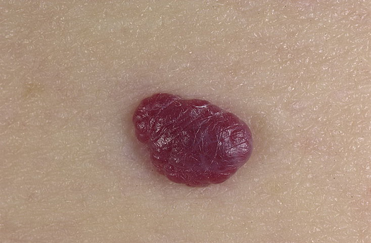
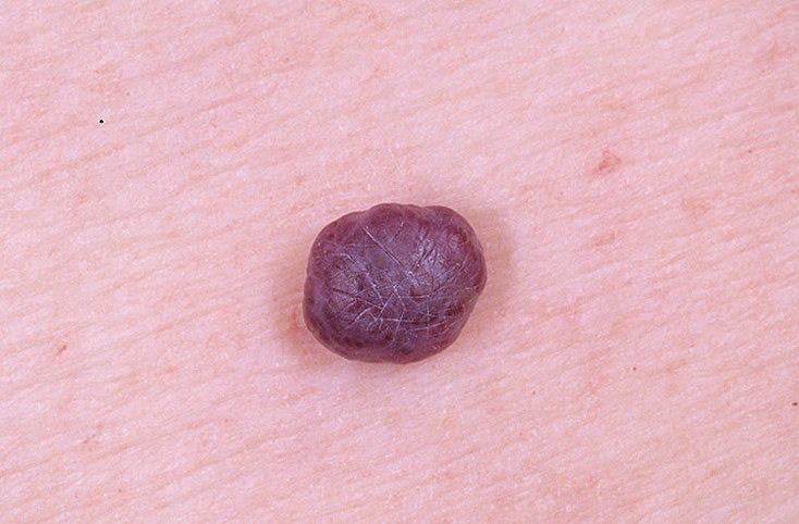
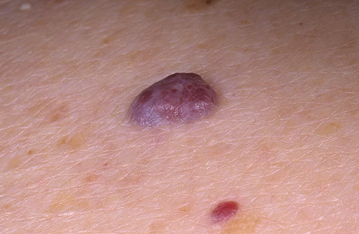
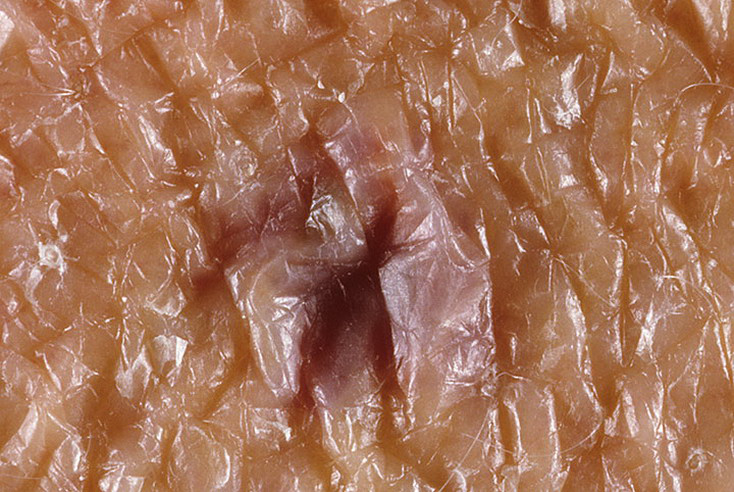
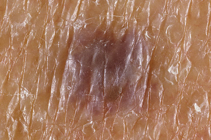
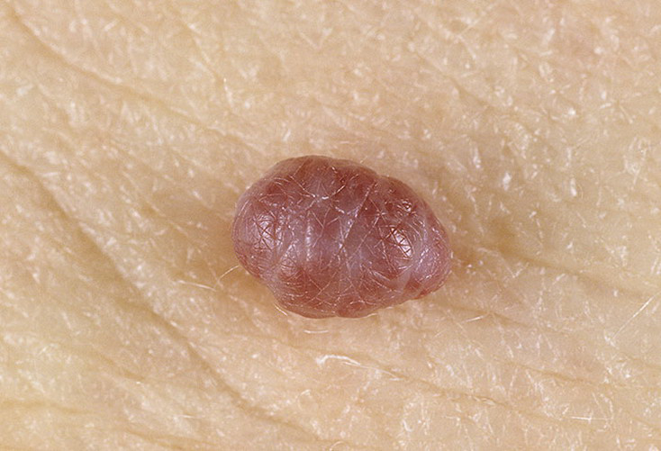
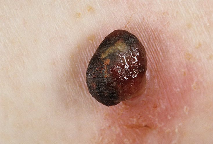
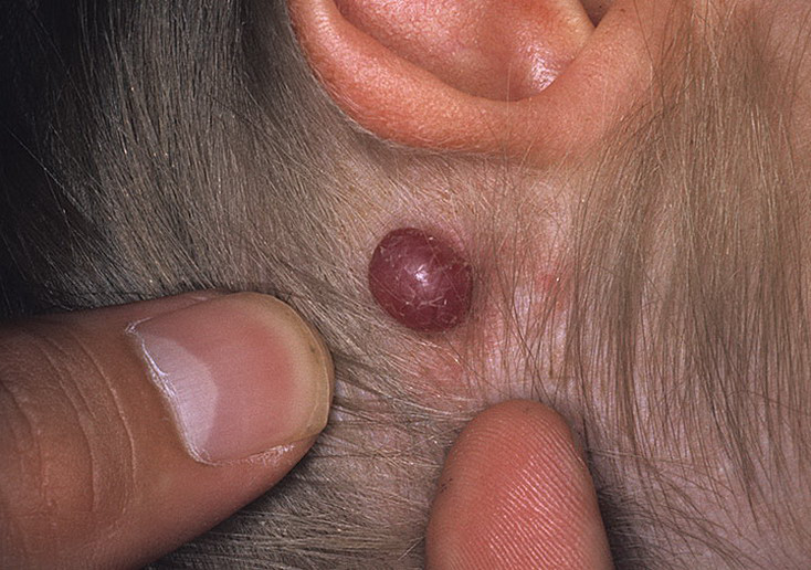
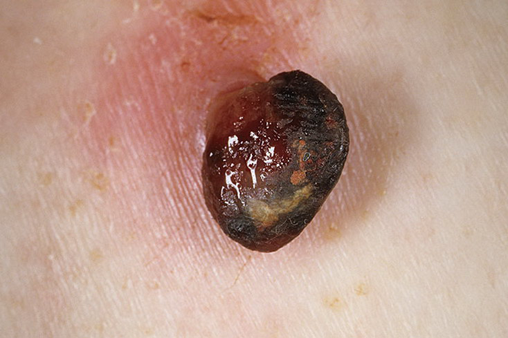
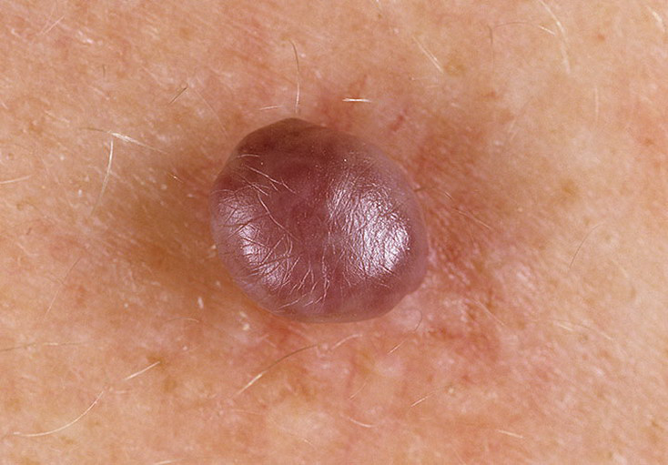
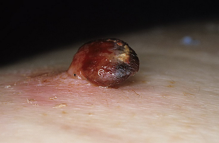
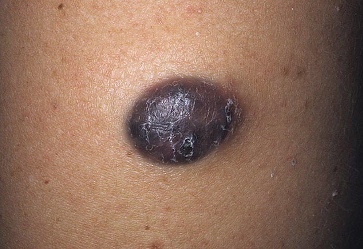
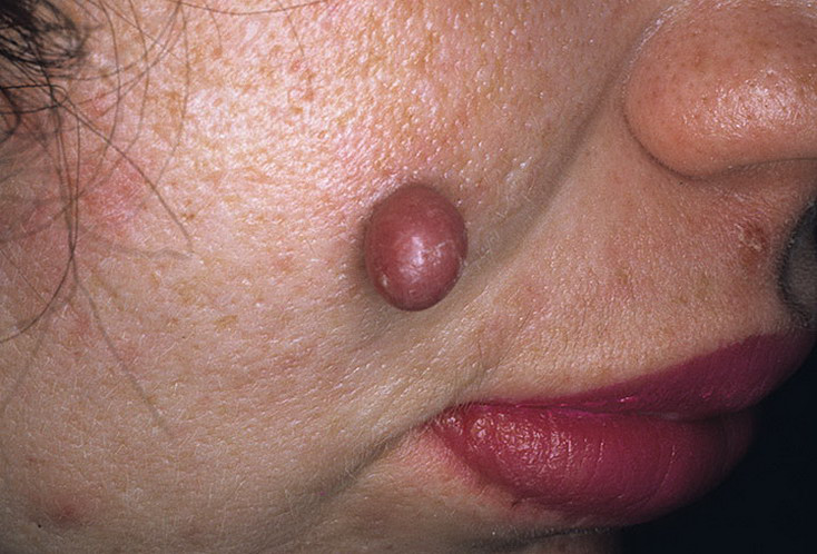
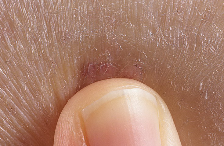
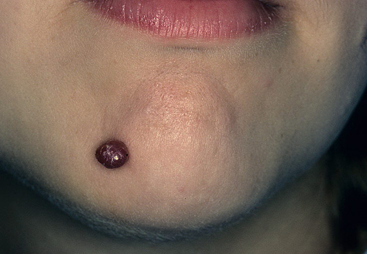
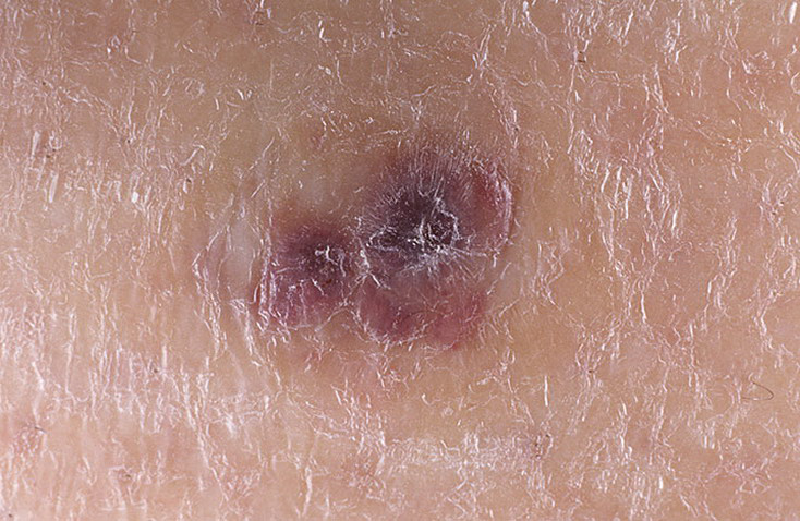
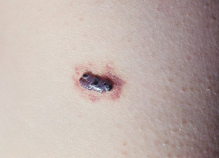
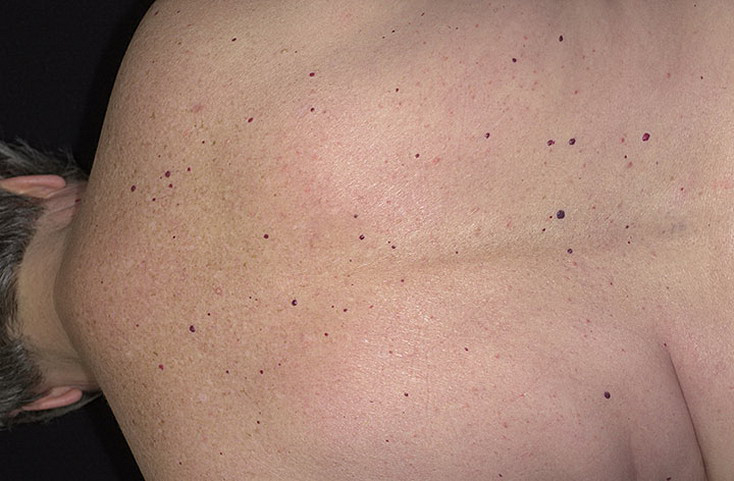
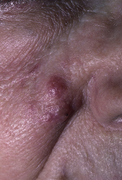
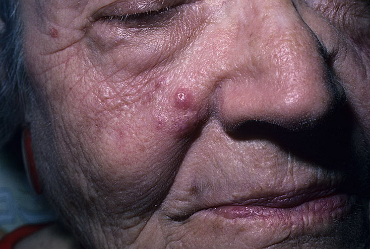
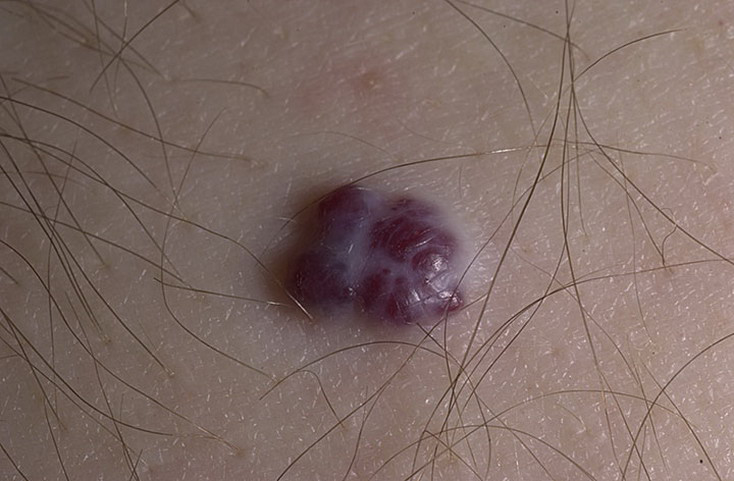
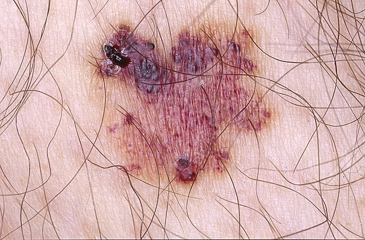
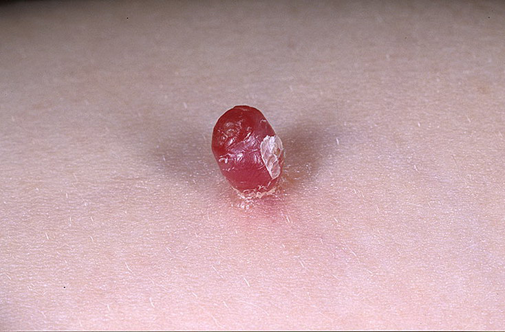
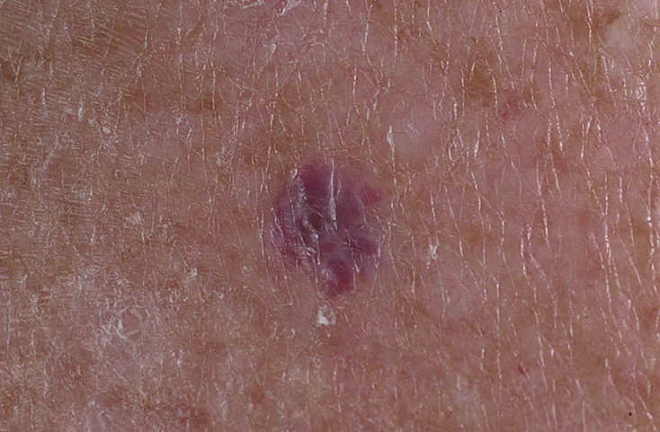
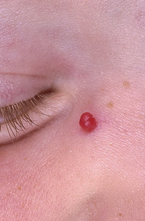
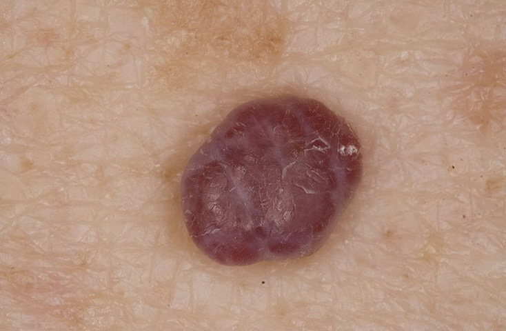
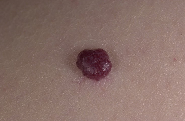
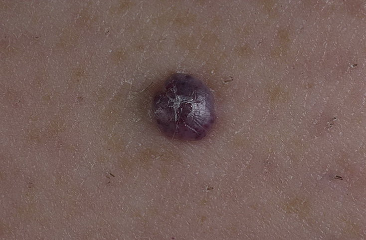
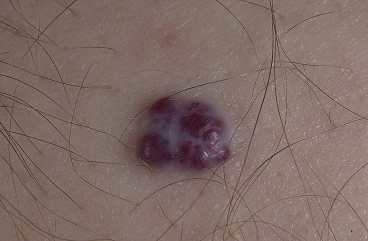
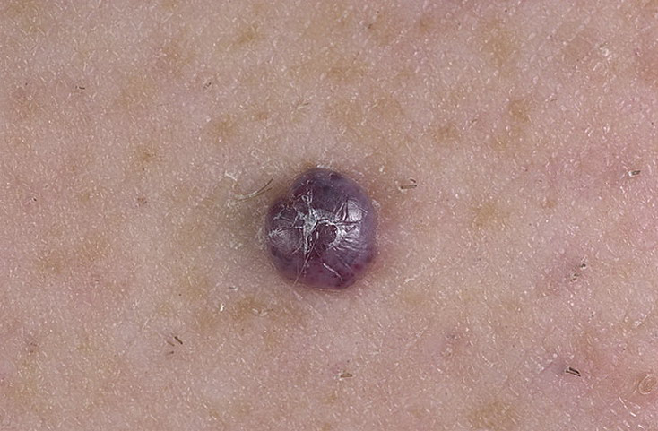
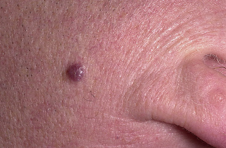
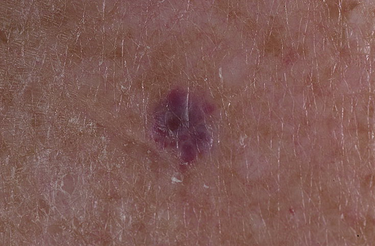
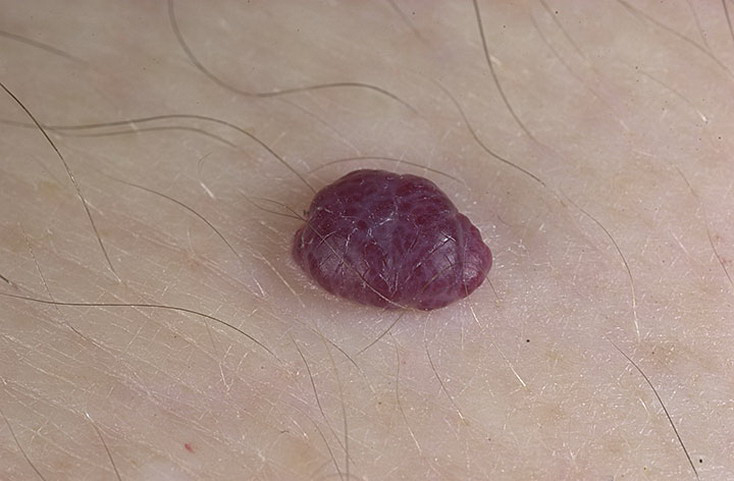
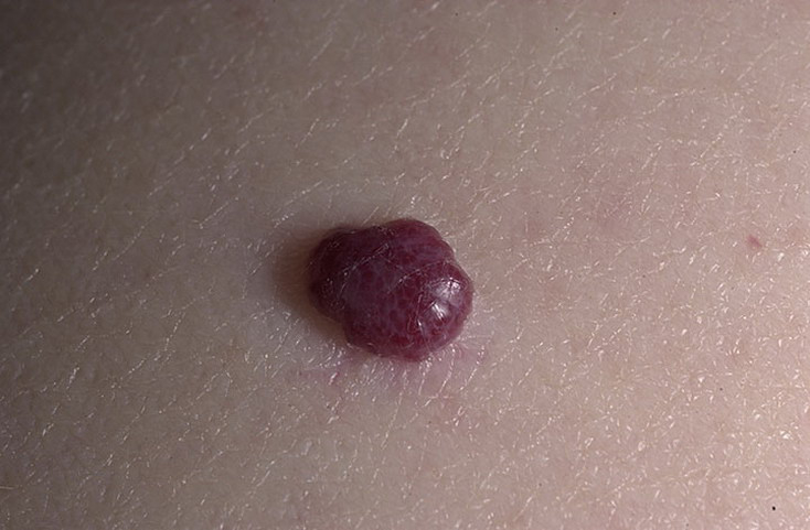
Hemangiomas occur most often in skin or subcutaneous tissue, and dermatologists, pediatricians, and primary care medical physicians typically treat these readily identifiable processes. One common example is the senile or cherry hemangioma, which is a benign, self-limited, small, red-purple skin papule seen in elderly patients. Another is the strawberry nevus, which is seen in approximately 0.5% of infants and spontaneously involutes in the vast majority of cases. Visceral hemangiomas are far less common but may have greater consequences when they result in organ dysfunction.
Orthopedists most commonly are called upon to treat hemangiomas of the deep soft tissues and bone. Skeletal muscle is the most common site for hemangioma of the deep soft tissue. Intramuscular hemangiomas may cause symptoms such as pain and swelling for which patients seek treatment. Hemangioma of bone may be symptomatic or may be purely an incidental finding. Most commonly, hemangiomas are localized to a single area, but multiple hemangiomas may occur in a single individual in a process known as hemangiomatosis.