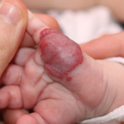
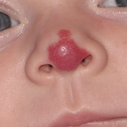
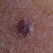
Cavernous Hemangioma in Newborns Pictures - 23 Photos & Images


Orbital cavernous hemangioma is a benign, slowly progressive vascular neoplasm of endothelial-lined spaces surrounded by a fibrous capsule. It most commonly presents in middle-aged adults (ages 20-40 years) and women are affected more than men. Its location is most often within the muscle cone, lateral to the optic nerve. Treatment is usually reserved for symptomatic patients (diplopia or visual disturbance) and includes surgical excision.
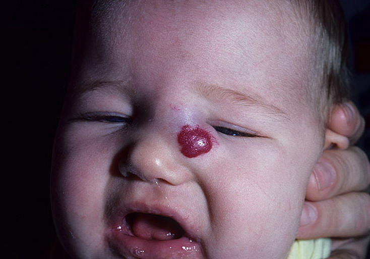
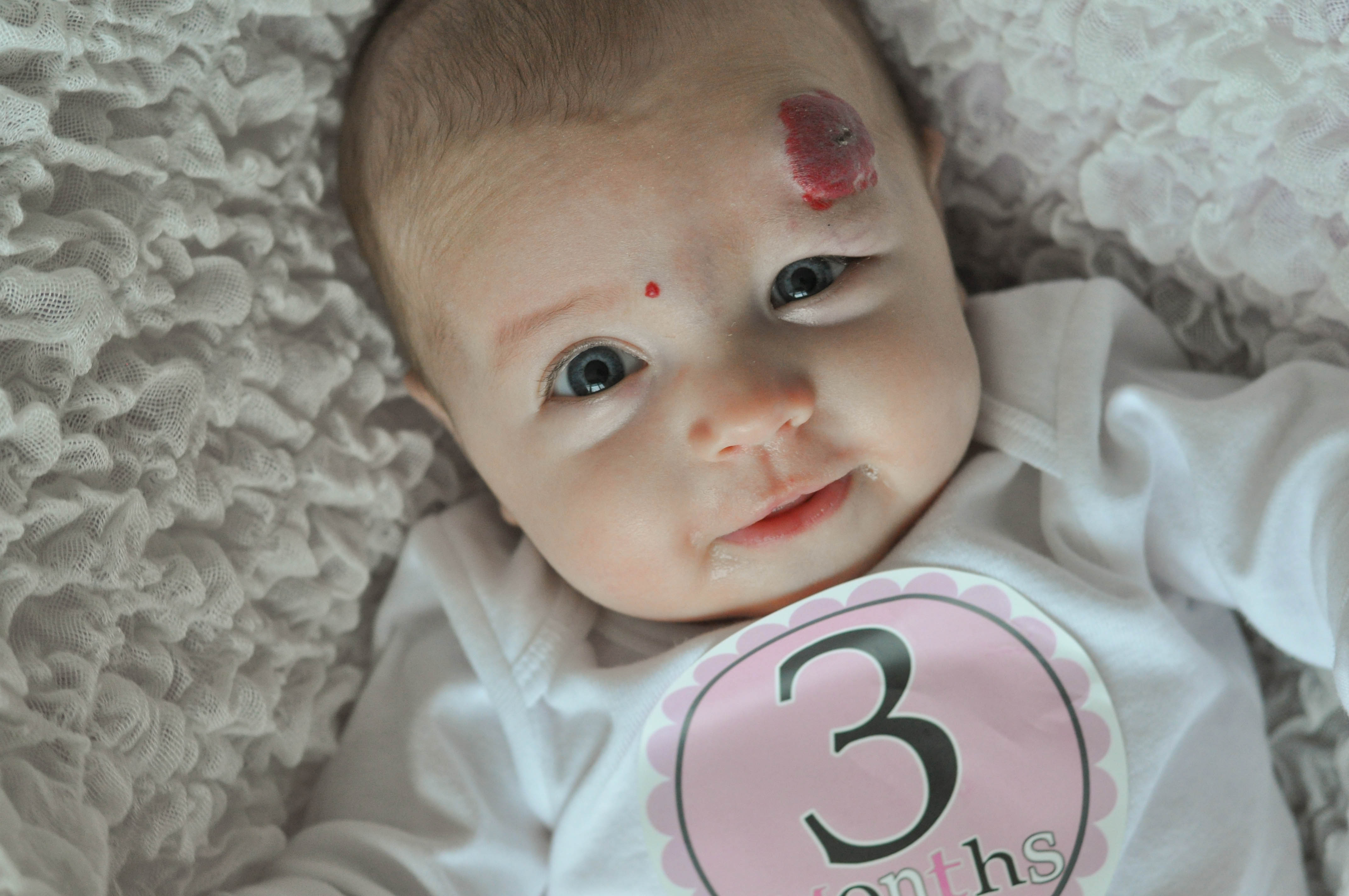
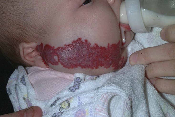
Cavernous hemangioma is the most common benign neoplasm of the orbit. It is considered a congenital abnormality. There is no evidence to suggest a heritance pattern. It is not a neoplasm in the usual sense, as it is not derived from a single cell, proliferating cell. Instead, cavernous hemangiomas are composed of a network of vascular channels separated by fibrous tissue stroma. Growth of the tumor is secondary to budding of vascular channels into surrounding tissue.
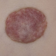
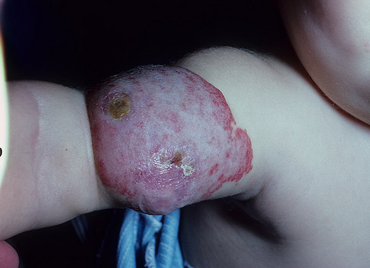
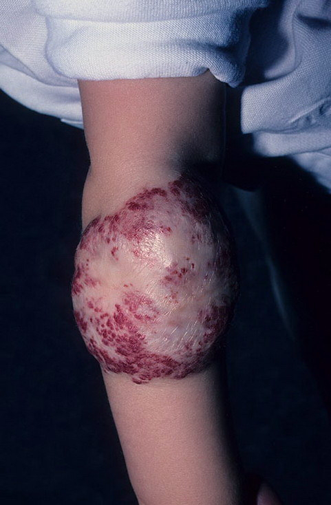
There are no established risk factors for cavernous hemangioma. However, pregnancy has been associated with accelerated growth of pre-existing cavernous hemangiomas. Cavernous hemangioma is an encapsulated nodular mass composed of dilated, cavernous vascular spaces separated by connective tissue stroma. Flattened endothelial cells line the vascular spaces, which are filled with blood. One to five layers of smooth muscle cells surround the vascular spaces. These histopathologic features may also be seen in lymphangioma.
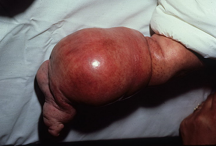
Cavernous hemangioma is a congenital abnormality that presents after sufficient growth causes cosmetic or visual disturbance. Growth of the tumor is a result of budding of the vascular channels into the surrounding soft tissue. It has been speculated that a localized, low-grade change in hemodynamics causes opening of new channels allowing for extension of the tumor into the surrounding interstitium. A fibrous capsule forms at the interface of the advancing tumor and the normal neighboring tissue. Surrounding soft tissue is displaced, compressed or occasionally incorporated into the tumor. Symptomatic visual impairment occurs as a result of involvement of the optic nerve, extraocular muscles or surrounding vasculature. Cavernous hemangioma is a congenital vascular neoplasm. There is no primary prevention for this disease entity
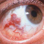
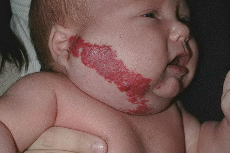
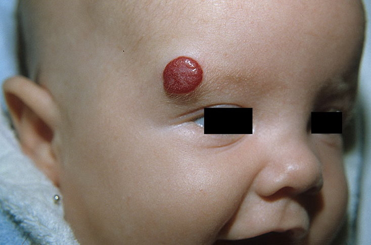
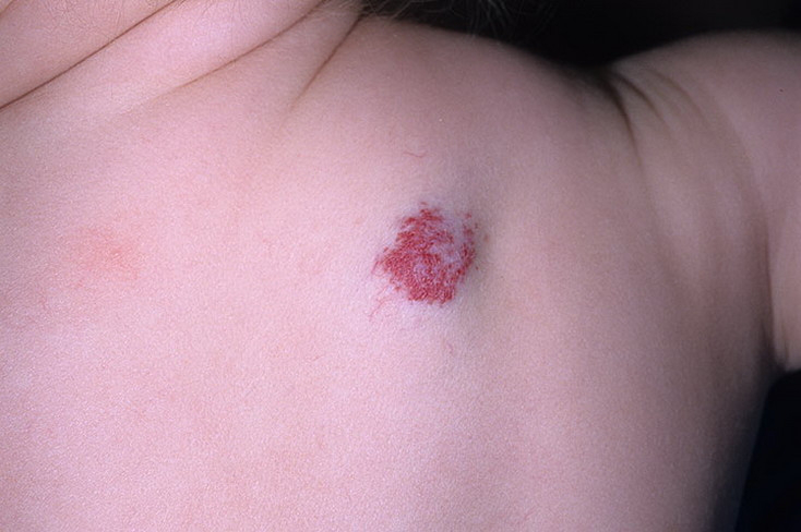
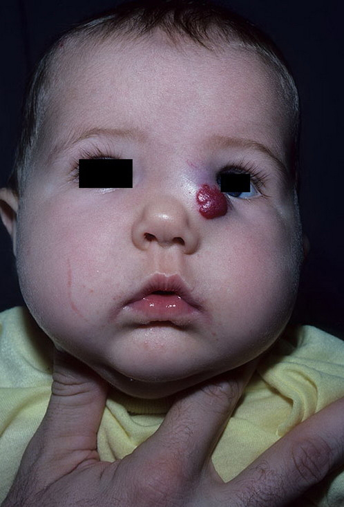
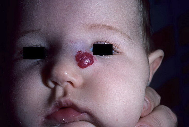
The diagnosis of cavernous hemangioma is suspected clinically and confirmed with orbital imaging, most commonly MRI. They most commonly present as solitary, unilateral lesions. However, multiple tumors have been reported, particularly in Blue Rubber Bleb Nevus Syndrome. Rarely, they may present simultaneously with a cavernous hemangioma of the brain. Cavernous hemangiomas have also been reported to present as an osseous lesion involving the orbital bones or as a tumor within the lacrimal gland.
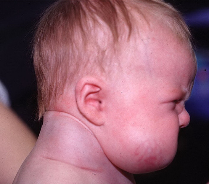
Depending on the size and location of the cavernous hemangioma, exam findings may range from normal to severe axial proptosis with poor vision, elevated intraocular pressure (IOP), motility defects and a relative afferent pupillary defect. Physical exam should include assessment of visual acuity, pupillary reaction,color vision, Hertel exophthalmometry, intraocular pressure, slit lamp exam with fluorescein and dilated fundus exam and visual fields in case of suspected optic nerve compression. A tumor of sufficient size may induce hyperopia or compress the optic nerve causing decreased visual acuity or an afferent pupillary defect. Hertel exophthalmometry will enable the examiner to appreciate small amounts of proptosis. Slit lamp exam with fluorescein instillation should be used to assess for surface irregularities that may occur as a result of incomplete lid closure from proptosis. Dilated fundus exam may reveal optic nerve swelling, choroidal folds or circumscribed compression of the globe.
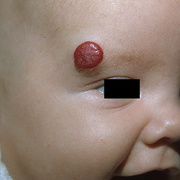
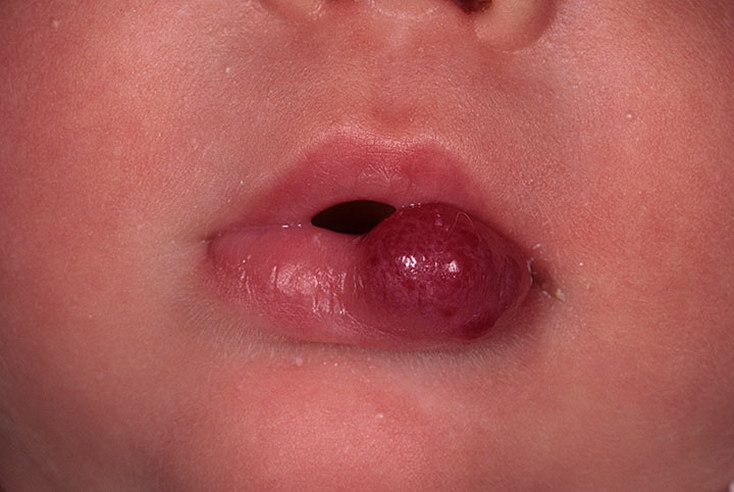
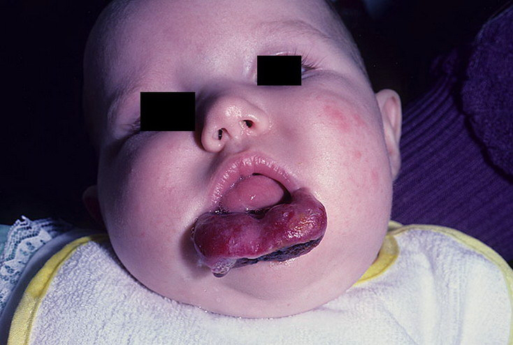
Patients with cavernous hemangiomas usually present with painless, progressive proptosis. As the tumor grows and involves the extraocular muscles, optic nerve and globe, patients will report double vision and decreased vision.
The diagnosis of cavernous hemangioma may be suspected clinically but is confirmed with orbital imaging. A presenting complaint of proptosis should always prompt a consideration of orbital imaging. Pupillary abnormalities elevated IOP, optic nerve swelling and choroidal folds should raise suspicion of a possible orbital mass.