Cavernous Hemangioma Pictures - 87 Photos & Images


Cavernous hemangioma, also called cavernous angioma, or cerebral cavernoma (when referring to presence in the brain) is a type of blood vessel malformation or hemangioma, where a collection of dilated blood vessels form a benign tumor. Because of this malformation, blood flow through the cavities, or caverns, is slow. Additionally, the cells that form the vessels do not form the necessary junctions with surrounding cells. Also, the structural support from the smooth muscle is hindered, causing leakage into the surrounding tissue. It is the leakage of blood, known as a hemorrhage from these vessels that causes a variety of symptoms known to be associated with this disease.
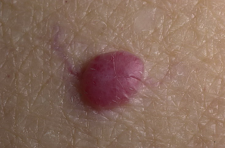
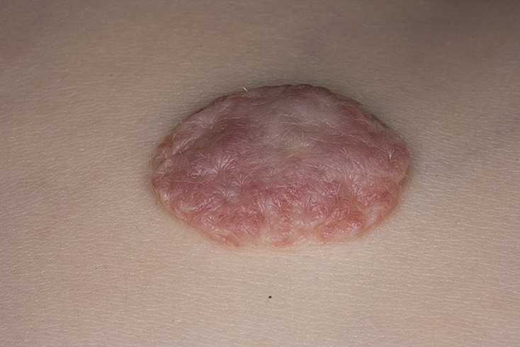
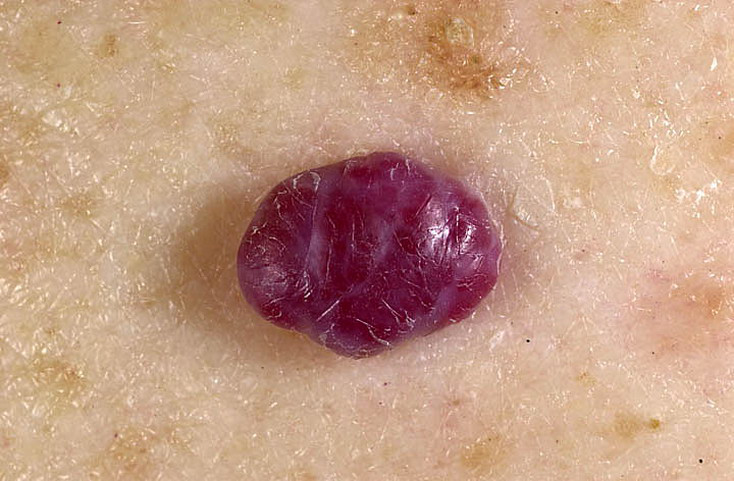
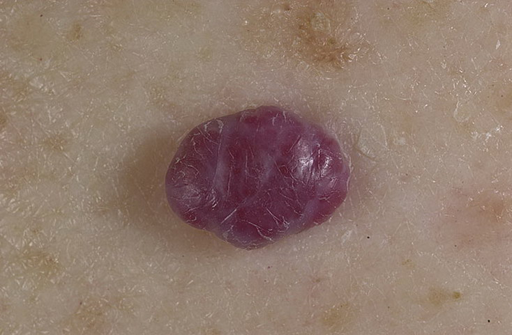
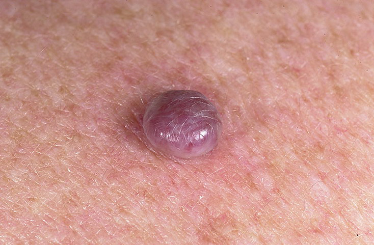
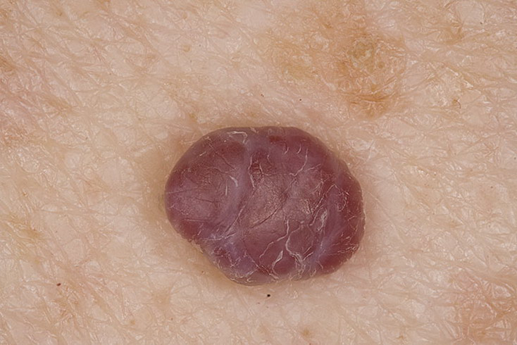
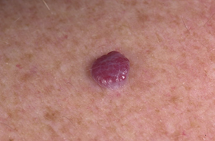
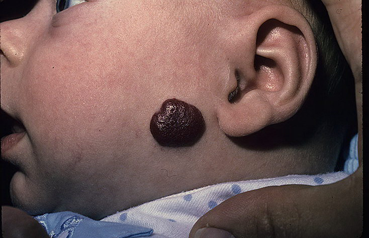
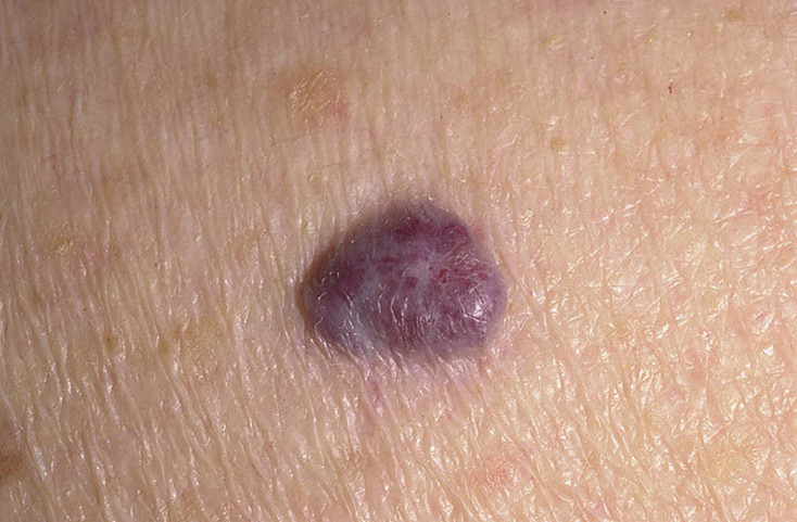
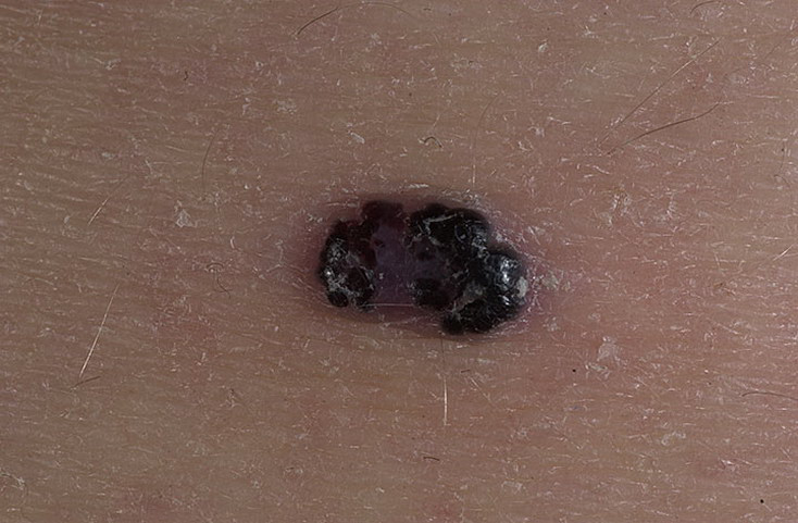
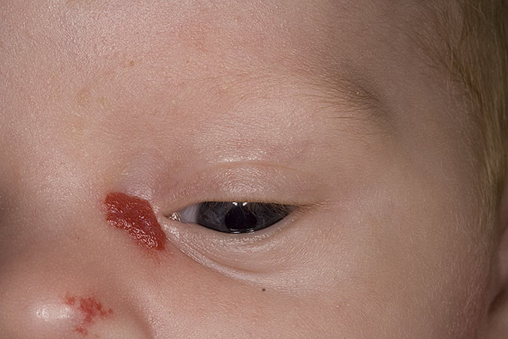
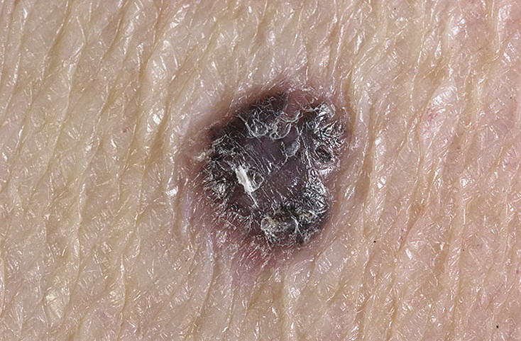
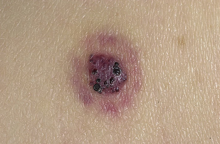
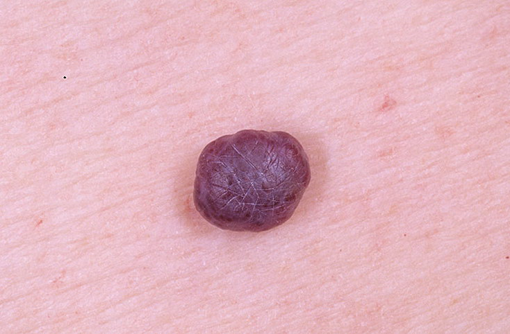
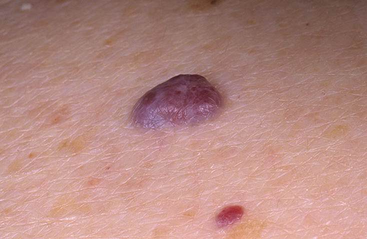
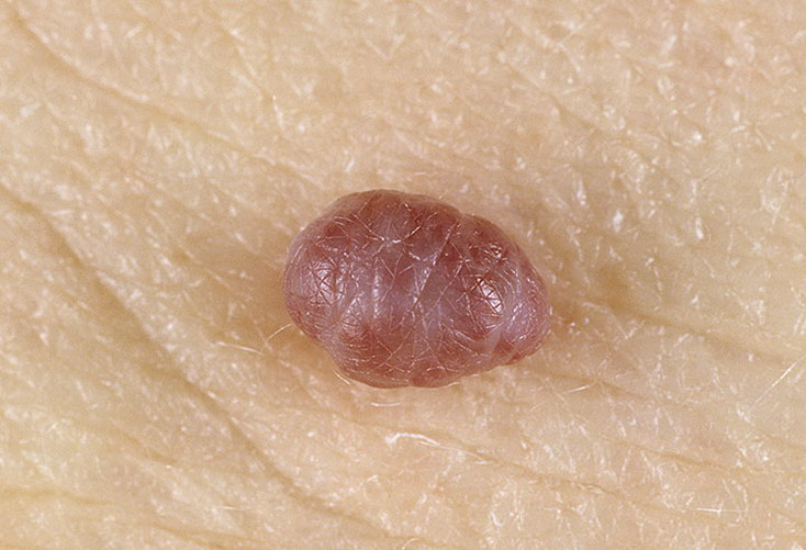
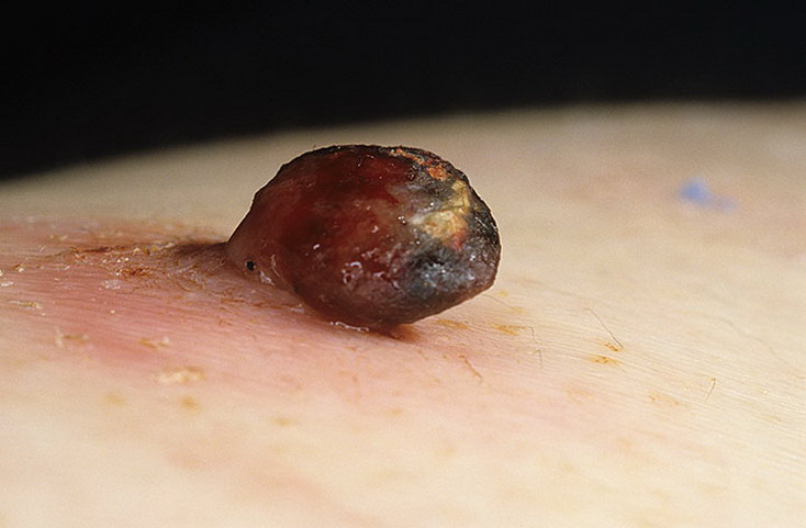
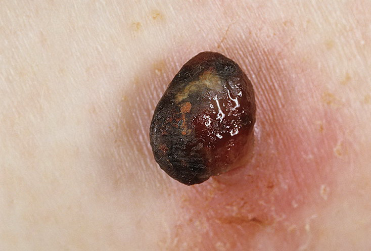
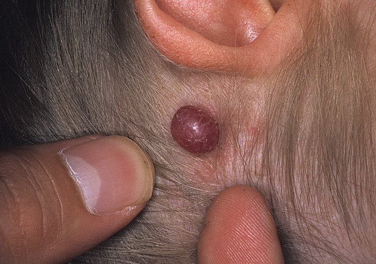
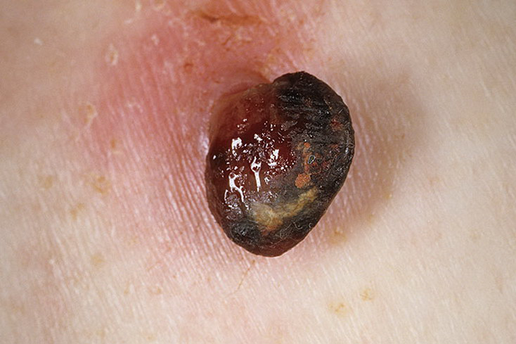
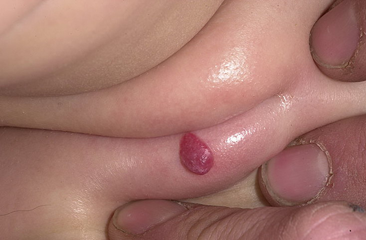
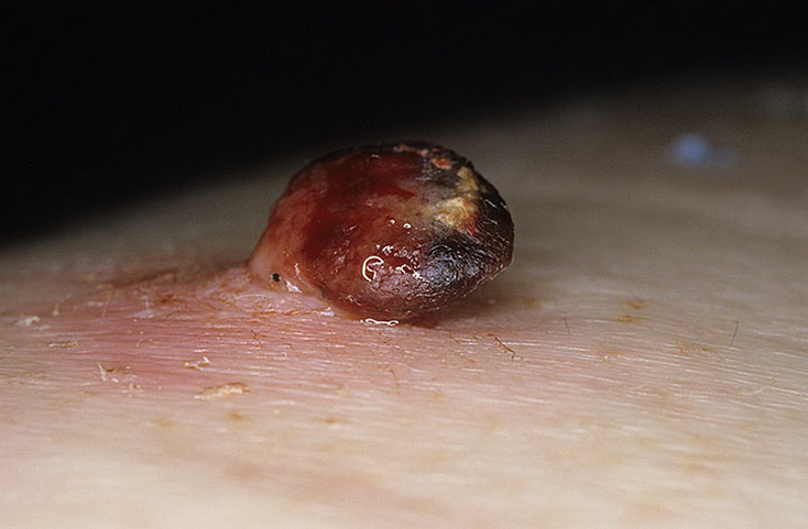
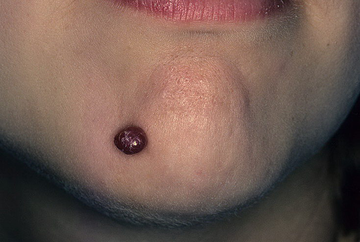
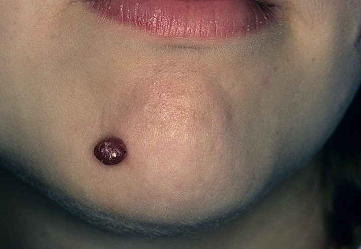
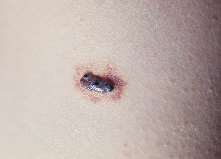
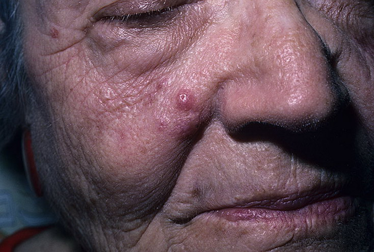
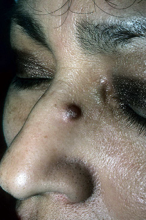
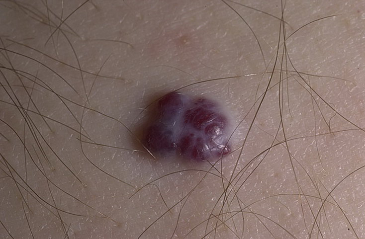
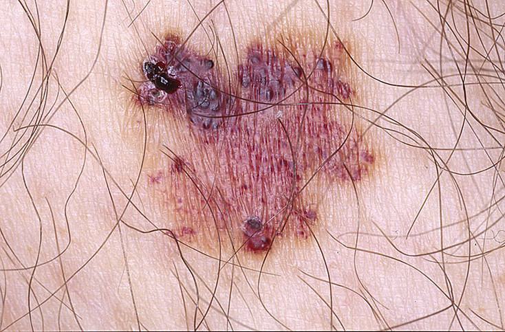
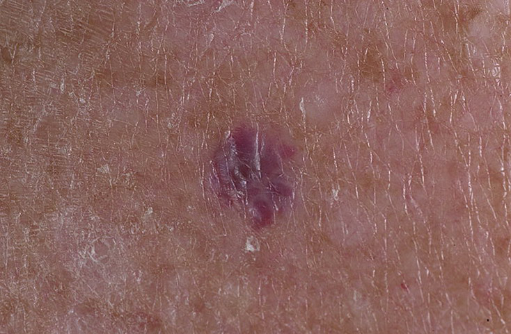
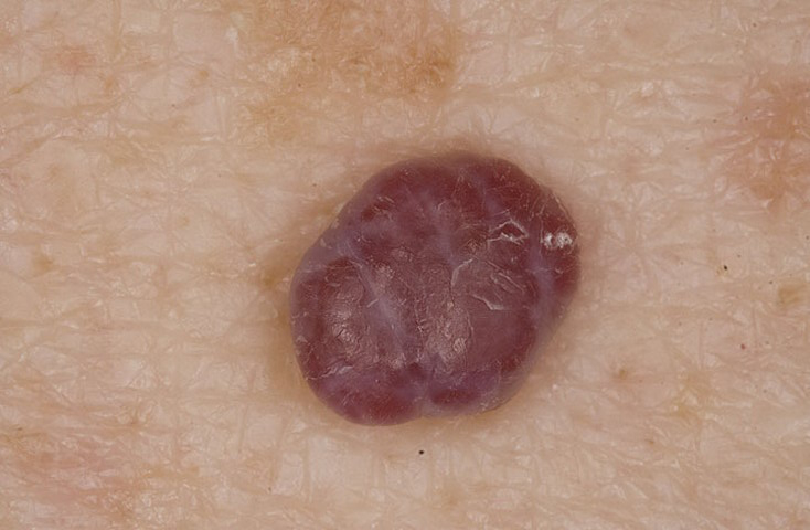
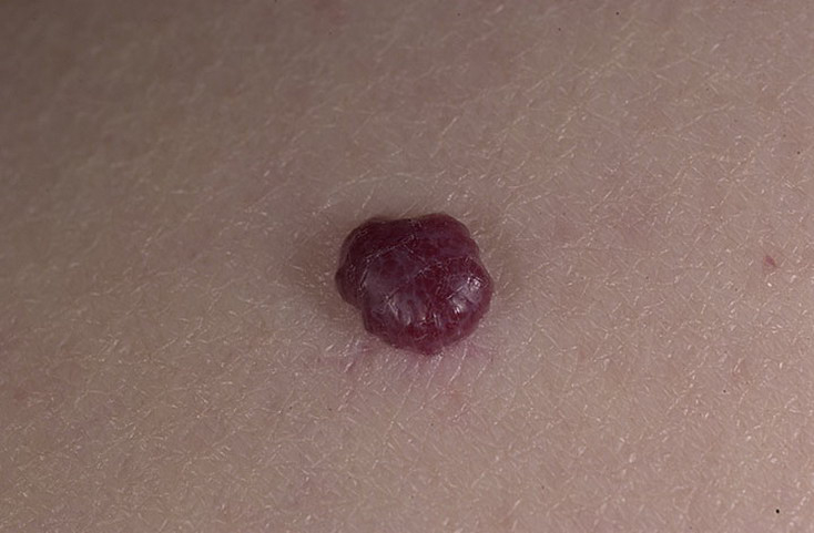
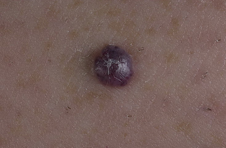
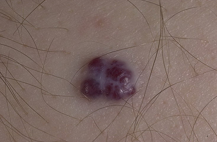
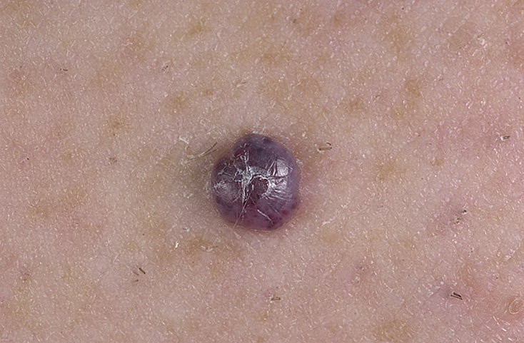
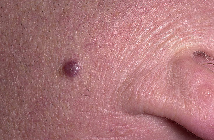
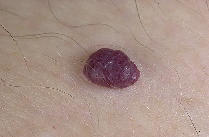
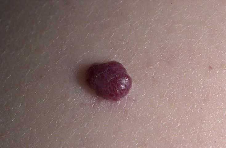
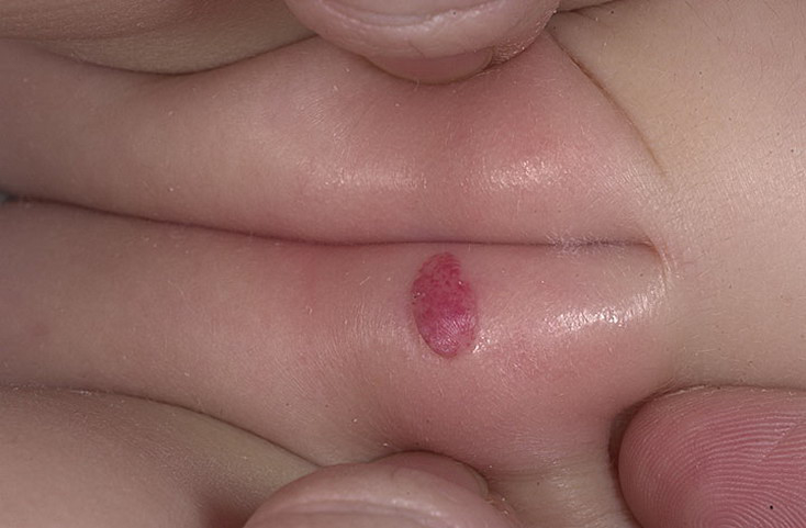
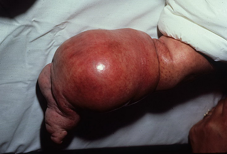
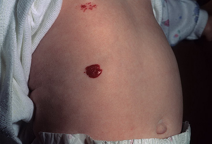
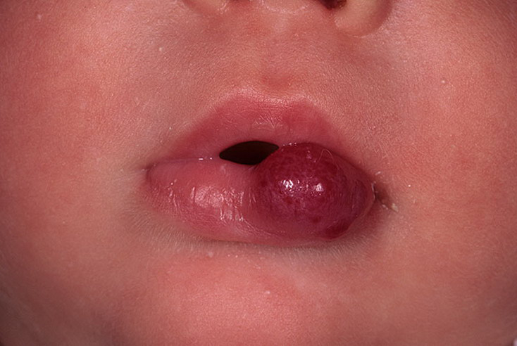
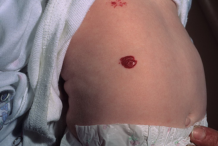
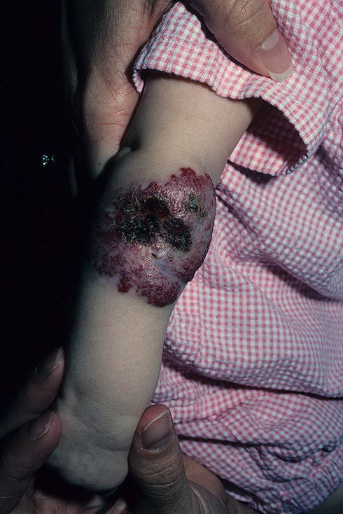
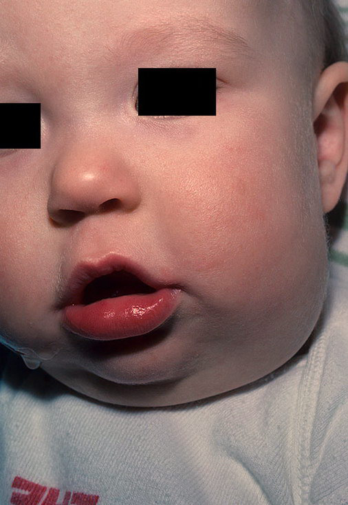
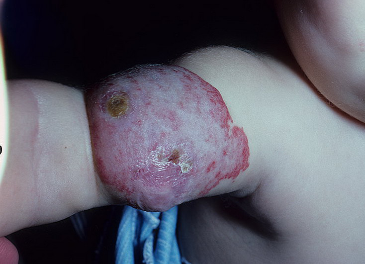
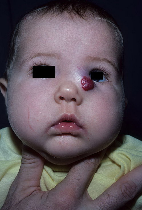
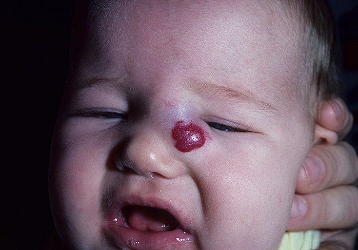
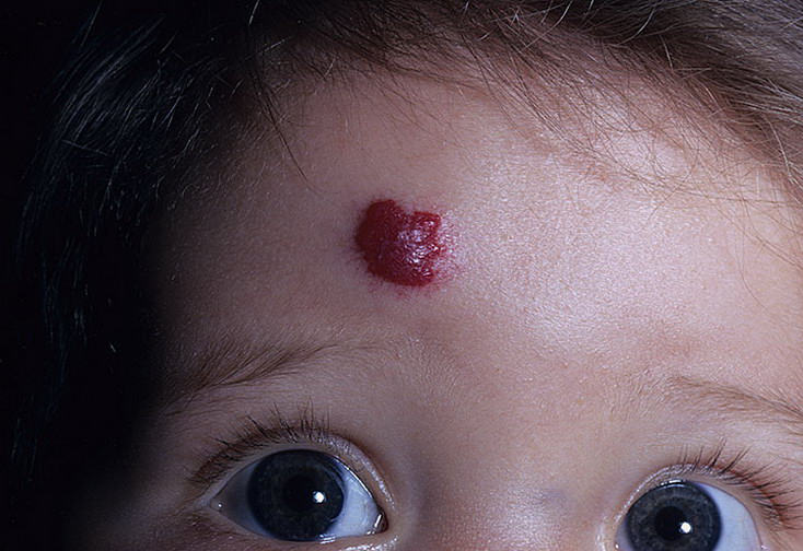
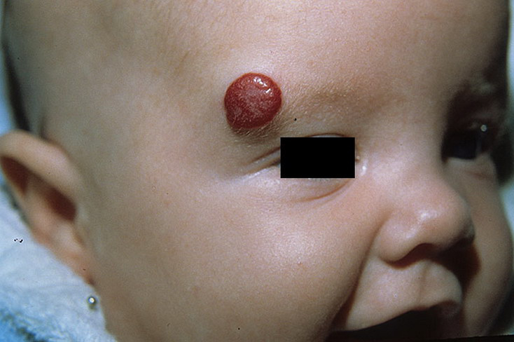
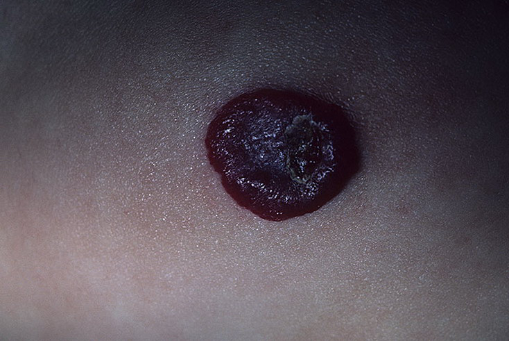
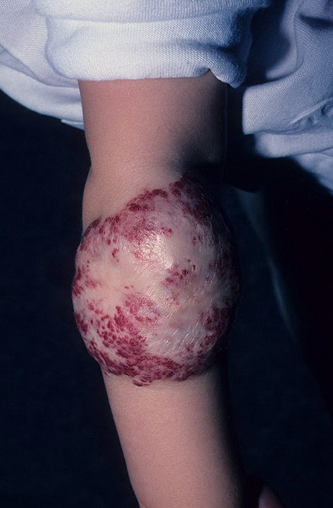
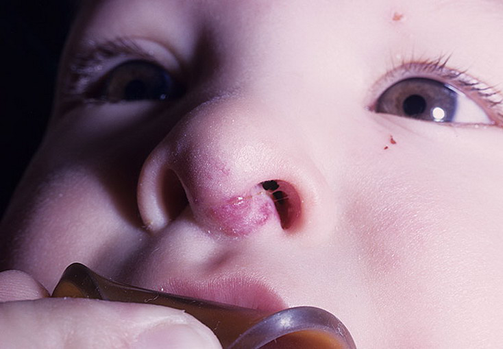
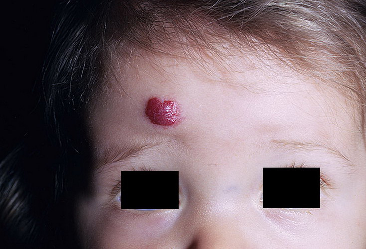
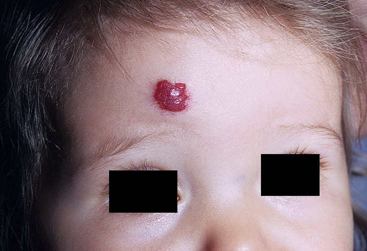
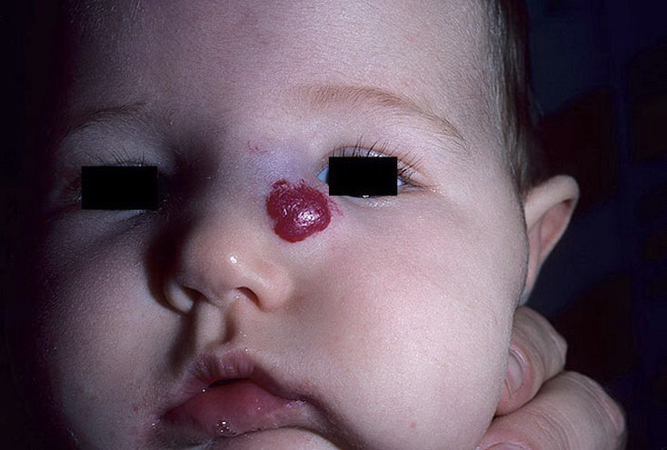
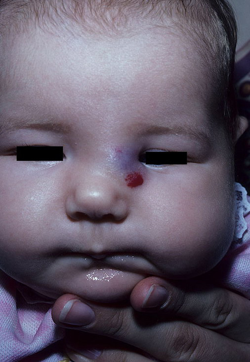
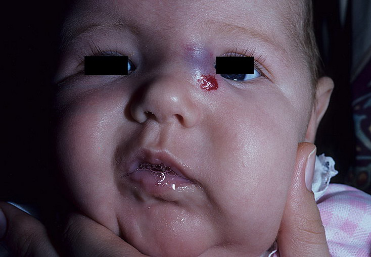
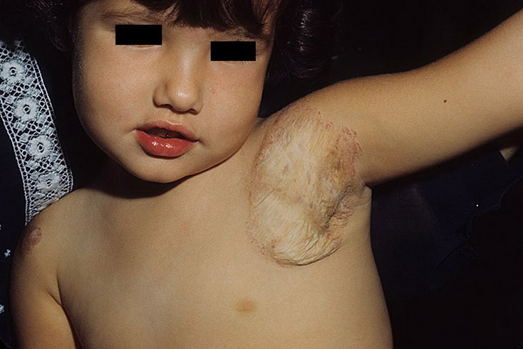
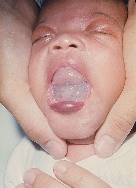
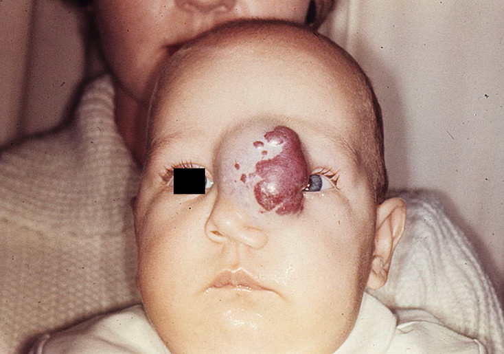
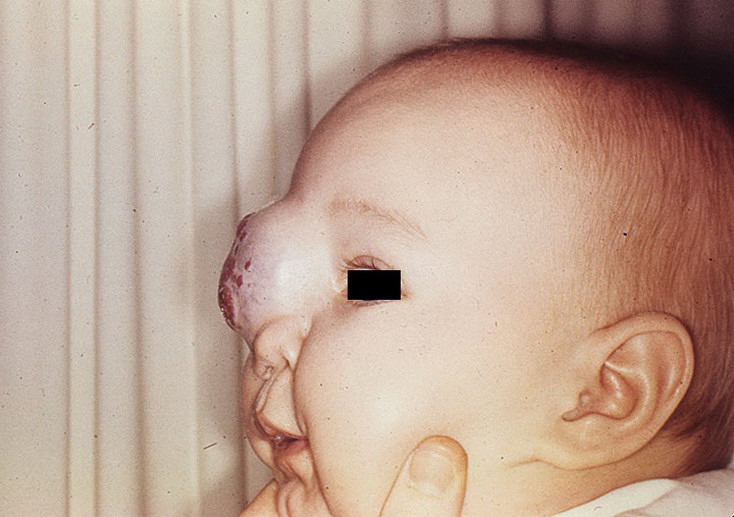
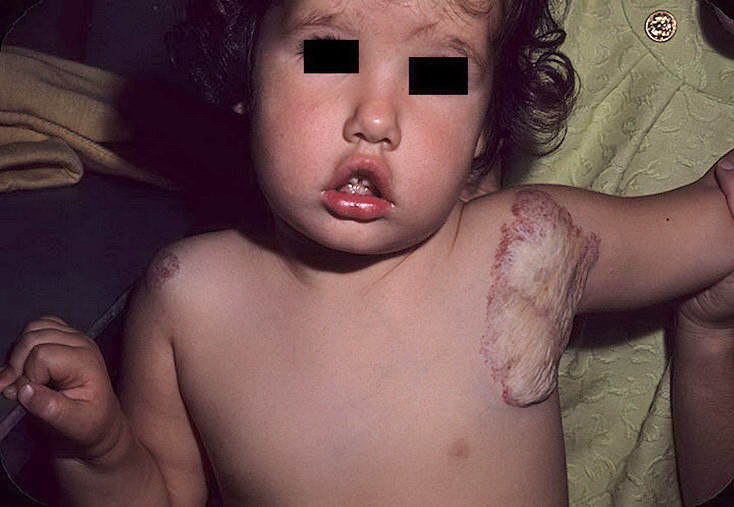
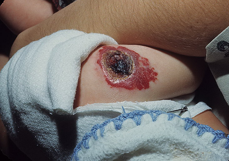
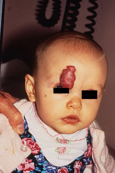
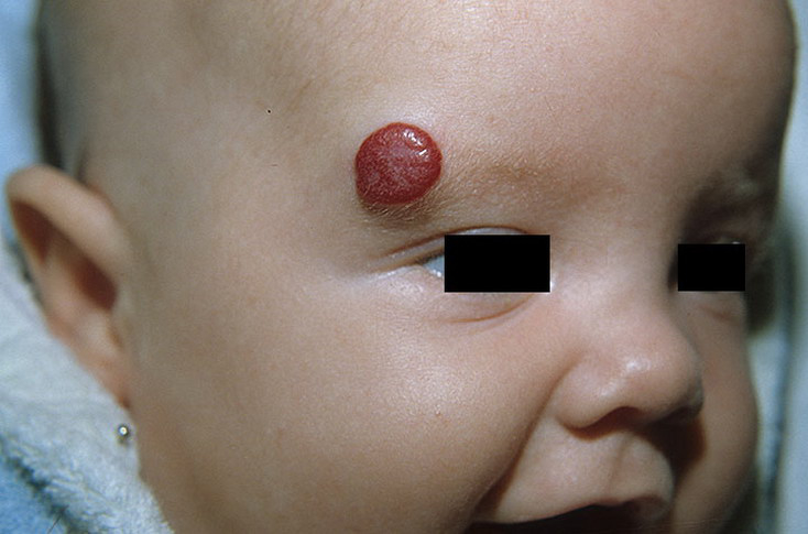
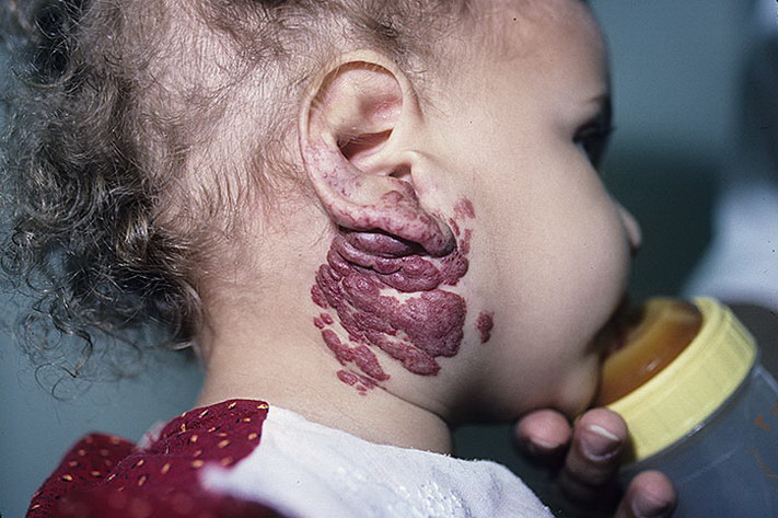
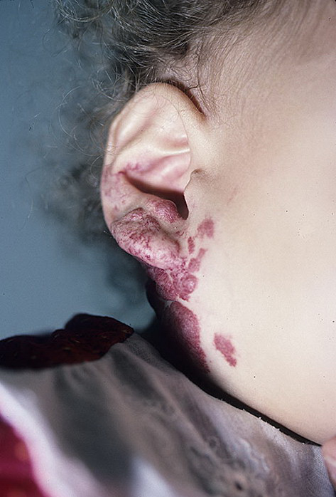
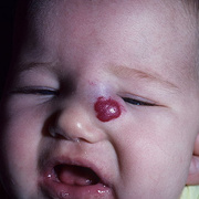
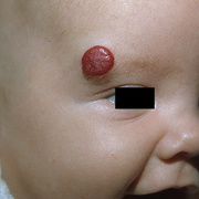
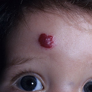
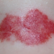
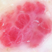
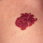
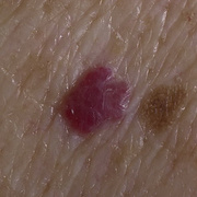
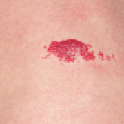
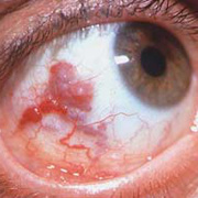
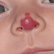
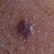
Cavernous hemangiomas can arise nearly anywhere in the body where there are blood vessels and are considered to be benign neoplasms (noncancerous tumors). They are often described as raspberry-like because of the bubble-like caverns. Unlike the capillary hemangiomas, cavernous ones can be disfiguring and do not tend to regress. Most cases of cavernomas are congenital, however they can develop over the course of a lifetime. While there is no definitive cause, research shows that genetic mutations result in the onset. Congenital hemangiomas that appear on the skin are known as either vascular or red birth marks.
Most patients are asymptomatic, but those who exhibit symptoms have a variety of treatment options that range from oral drugs to surgery depending on the malformation severity in order to decrease the rapid cell division or remove the lesion all together.
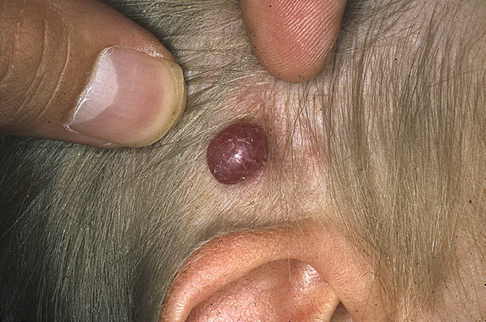
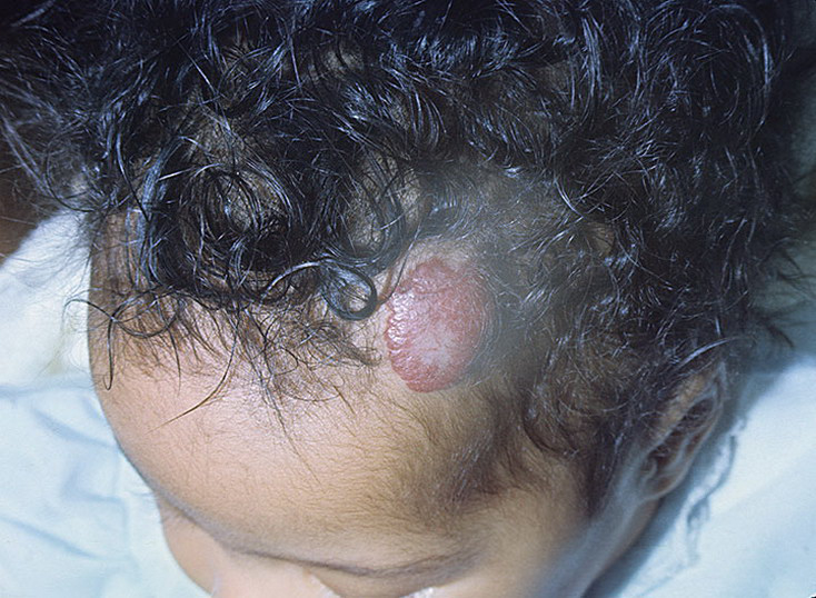
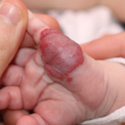
Small hemangioma on the scalp of a two-year-old female. Since the mid 19th century, pathologists have come across cases of cavernomas by looking at brain tissue under a microscope. Still most symptomatic cases were inaccurately diagnosed with other neurological diseases such as multiple sclerosis. When MRI was developed in the 1980s, the number of diagnosed cases of cavernous hemangioma increased.
My sister had what I know believe was cavernous hemangiona. She began having seizures in her 20's and continued having seizures until she died at the age of 37 3 days after 8.5 hrs of brain suregery in 1967. She was 8 mos pregnant with her 3rd child a few years before she died. She had a grand mal seizure and the baby separated from the placenta and died. How far we have come since then. The drs told us she has a mass of blood vesssels or capilairies in her brain, near the brain stem. As far as I know, no oither obvious symptoms were present. She often had double vision after a seizure. This information i have been reading about this disorder made me think about her life and how it was shortened. Her daughter, who is now in her eary 60's has been diagnosed with CH but no real symptoms that i know of. My sister's name was Roberta Schacht in Colorado.