Hemangioma Eyes Pictures - 3 Photos & Images

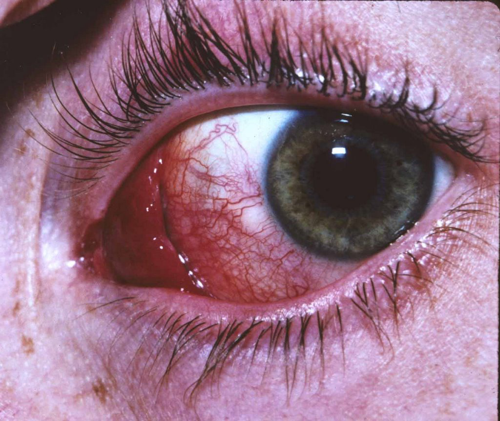
Hemangiomas typically grow within the blood vessel layer beneath the retina called the choroid. If they are located in the macula (center of vision) or they leak fluid (which causes a retinal detachment or cystic changes in the retina), they can affect your vision. Many choroidal hemangiomas never grow or leak fluid and may be observed without treatment. Choroidal hemangiomas are not cancers and never metastasize.
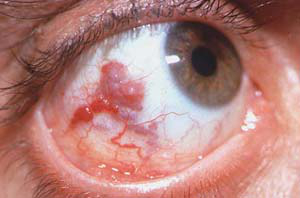
Choroidal hemangioma are typically reddish to orange. Choroidal hemangioma can have areas of increased pigmentation which (in those cases) can make them difficult to differentiate from choroidal melanomas. Choroidal hemangiomas can cause far-sightedness (hyperopia), distorted vision (metamorphopsia), flashing lights, or blurred vision. Many choroidal hemangioma cause no symptoms at all, and are found on routine dilated eye examinations (ophthalmoscopy).
Intraocular photography and angiography: Eye-care specialists perform studies of the blood vessels in the eye with synthetic organic dyes called fluorescein or indocyanine green. The dyes are injected into the arm or hand and travel to the blood vessels inside the eye. If a tumor is in the eye, we can see specific characteristics of its circulation which can help us differentiate between it and other types of tumors. Choroidal hemangioma often have a unique pattern of circulation where the large blood vessels produce a "COARSE VASCULAR PATTERN."