Nodular Melanoma Pictures - 27 Photos & Images


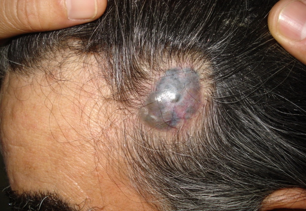


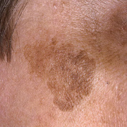
Nodular melanoma may arise on any site, but is most common on exposed areas of the head and neck.
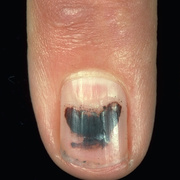
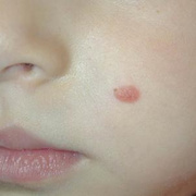
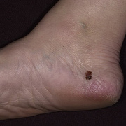
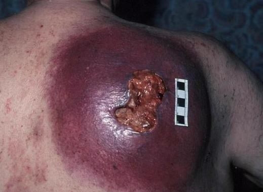
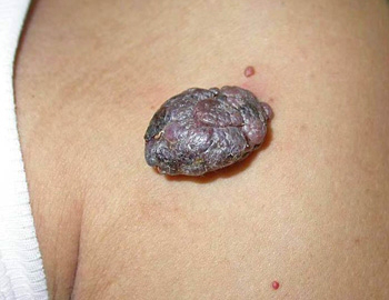
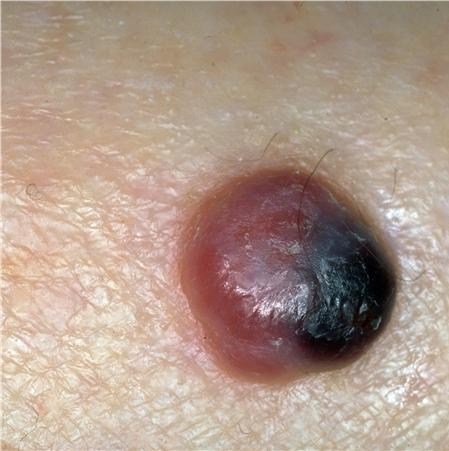
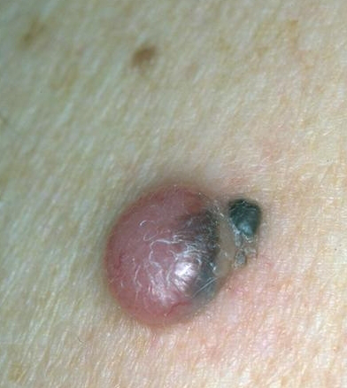
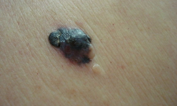
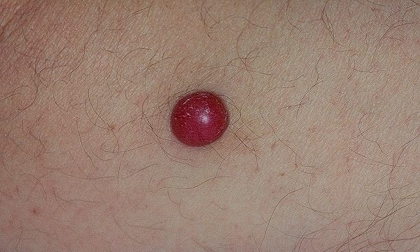
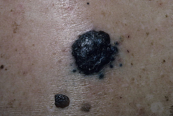
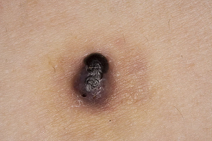
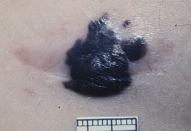
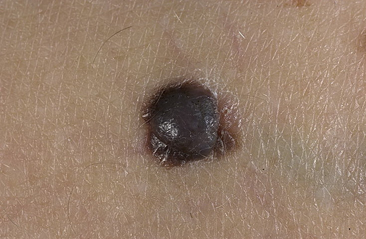
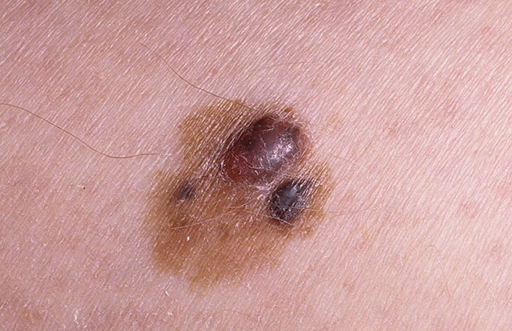
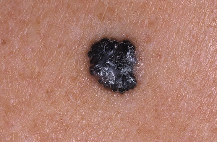
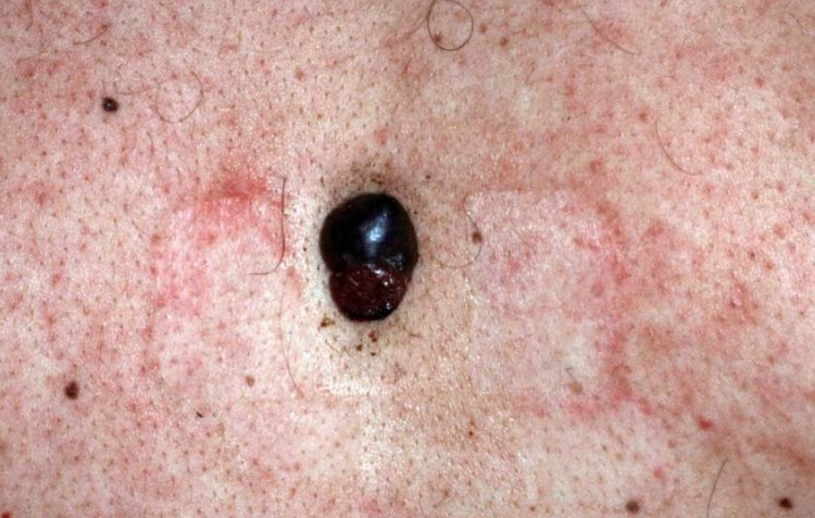
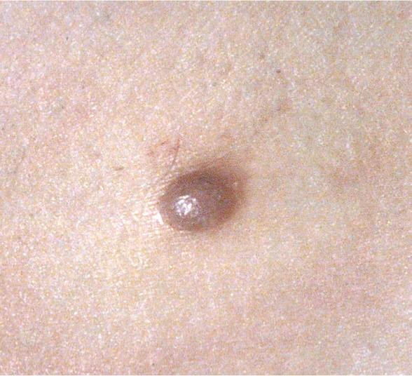
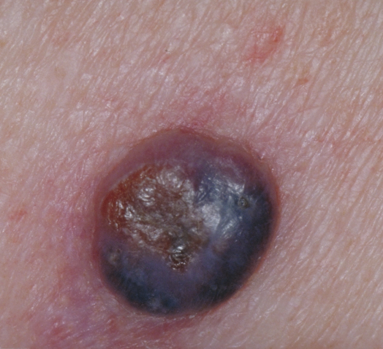
Nodular melanoma presents as a rapidly enlarging lump (over several weeks to months). The characteristics of nodular melanoma include:
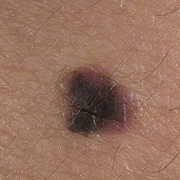
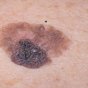
Larger size than most moles – >6 mm and often a centimetre or more in diameter at diagnosis
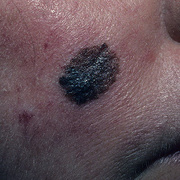
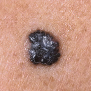
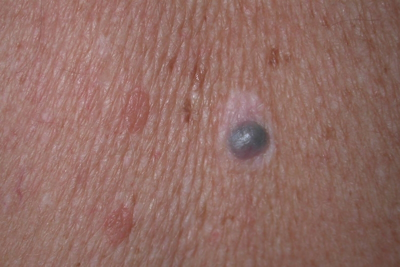
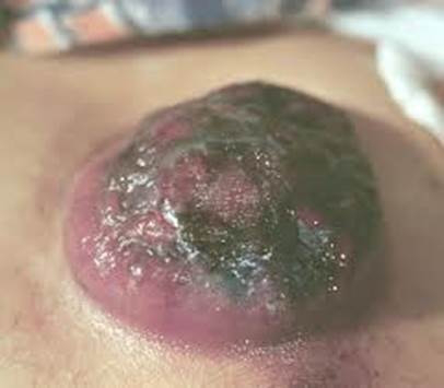
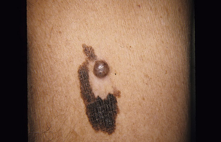
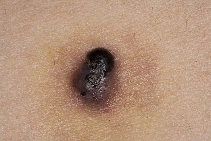
Dome-shaped, often symmetrical firm lump
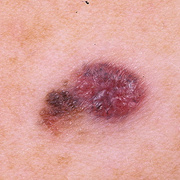
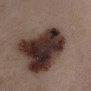
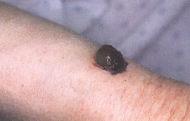
Single colour or variable pigmentation – most often black, red or skin coloured
Smooth, rough, crusted or warty surface
Ulceration or bleeding
Itching or stinging
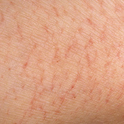
One-third of nodular melanomas are not pigmented. They lack the ABCD melanoma warning signs. (Asymmetry, Border irregularity, Colour variation, large Diameter).
Nodular melanoma is due to the development of malignant pigment cells (melanocytes) along the basal layer of the epidermis. These cells may occasionally arise from an existing melanocytic naevus (about 3%) but commonly arise within another type of melanoma or in previously normal-appearing skin. What triggers the melanocytes to become malignant is unknown, but it is likely to be a series of changes to the DNA. NRAS mutations are often found in patients with nodular melanomas.
It is essential to diagnose nodular melanoma accurately. Clinical diagnosis is aided by dermoscopy and skin biopsy (usually excision biopsy). Those with melanoma that is more than 1 mm thick may be advised to have lymph node biopsy, imaging studies and blood tests.