Bowenoid Papulosis Pictures - 20 Photos & Images
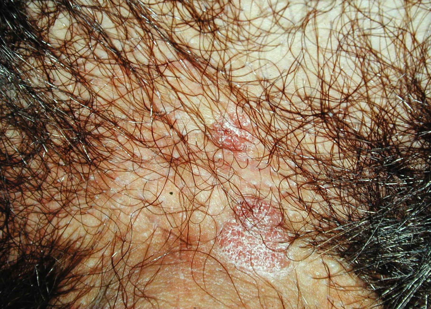
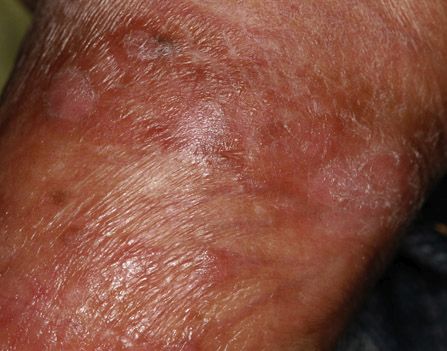
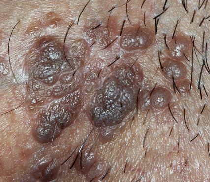

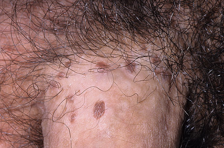
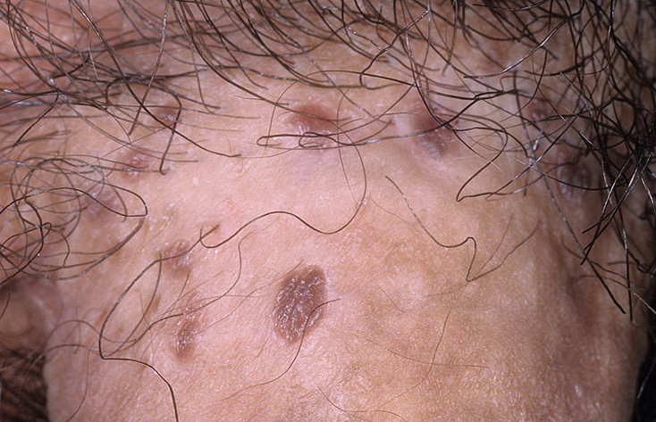
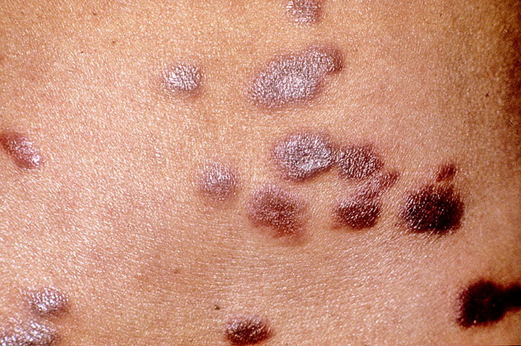
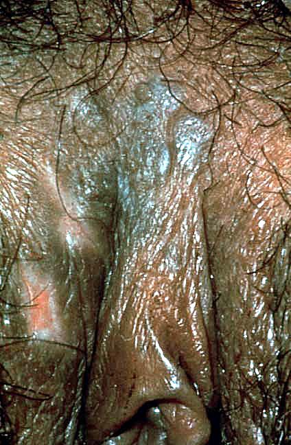
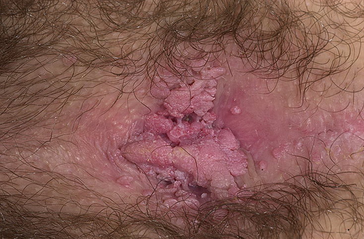
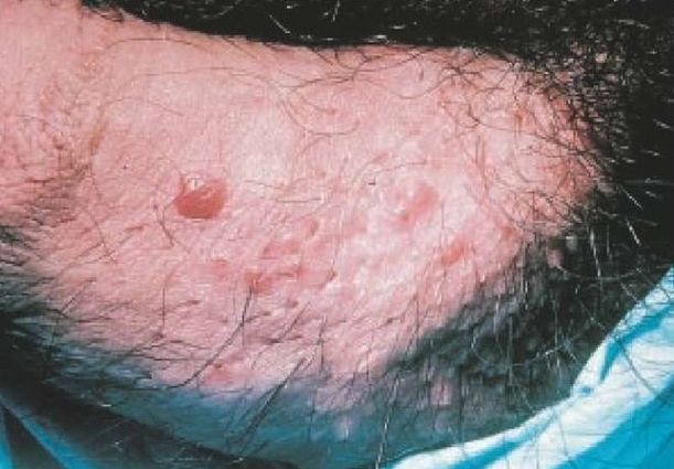

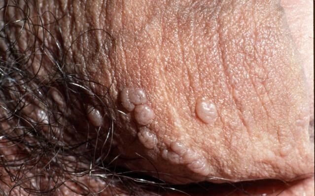
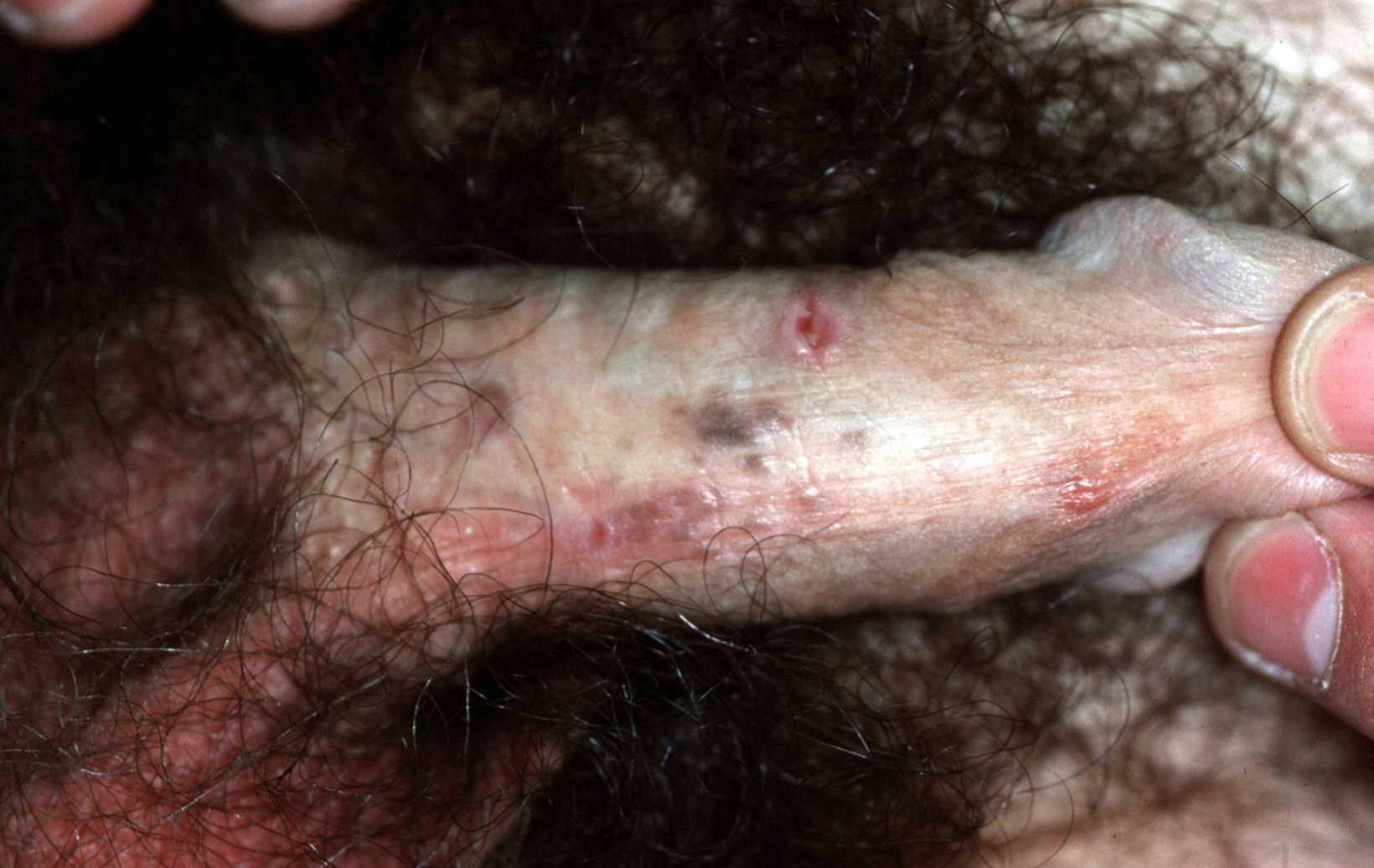


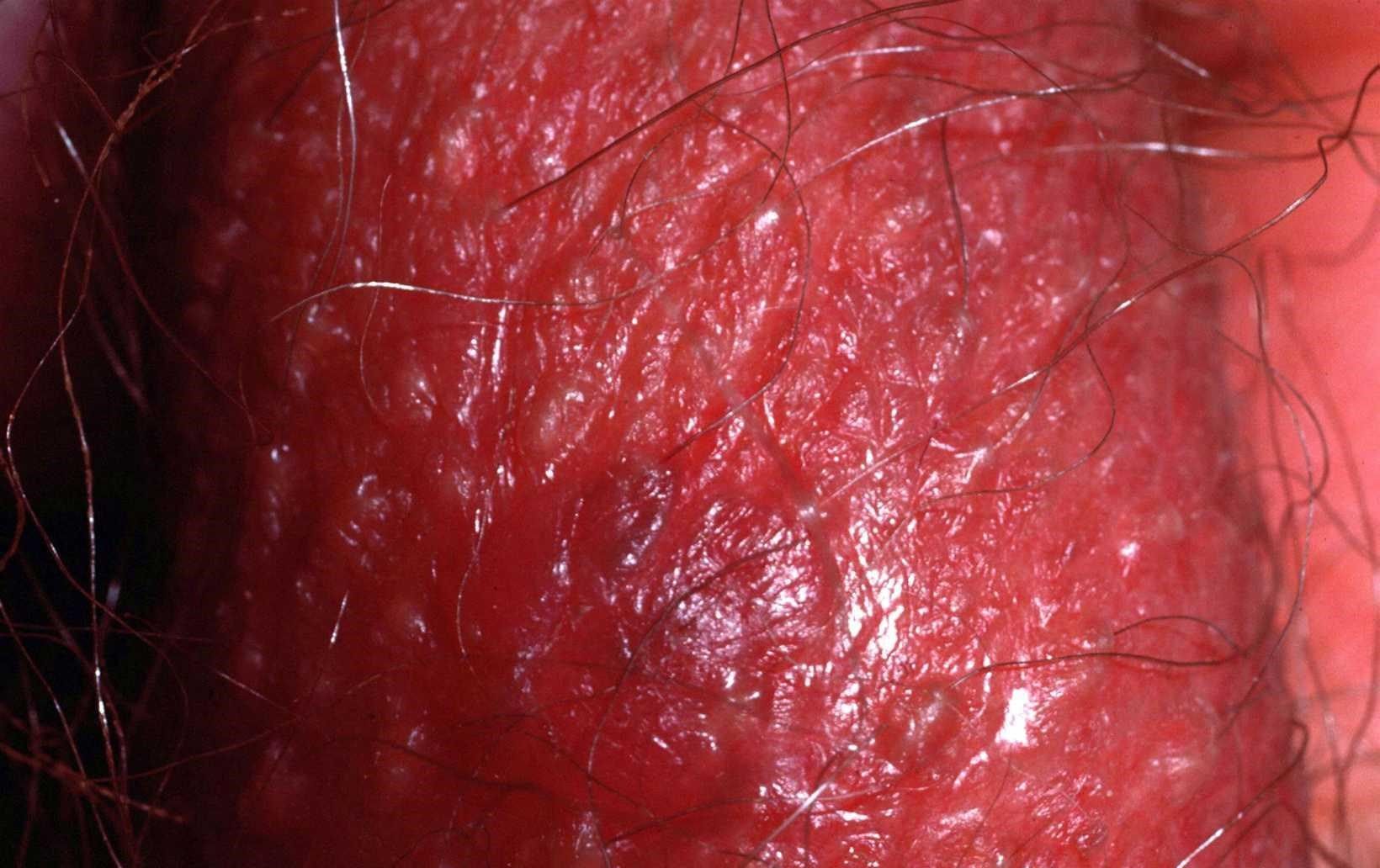
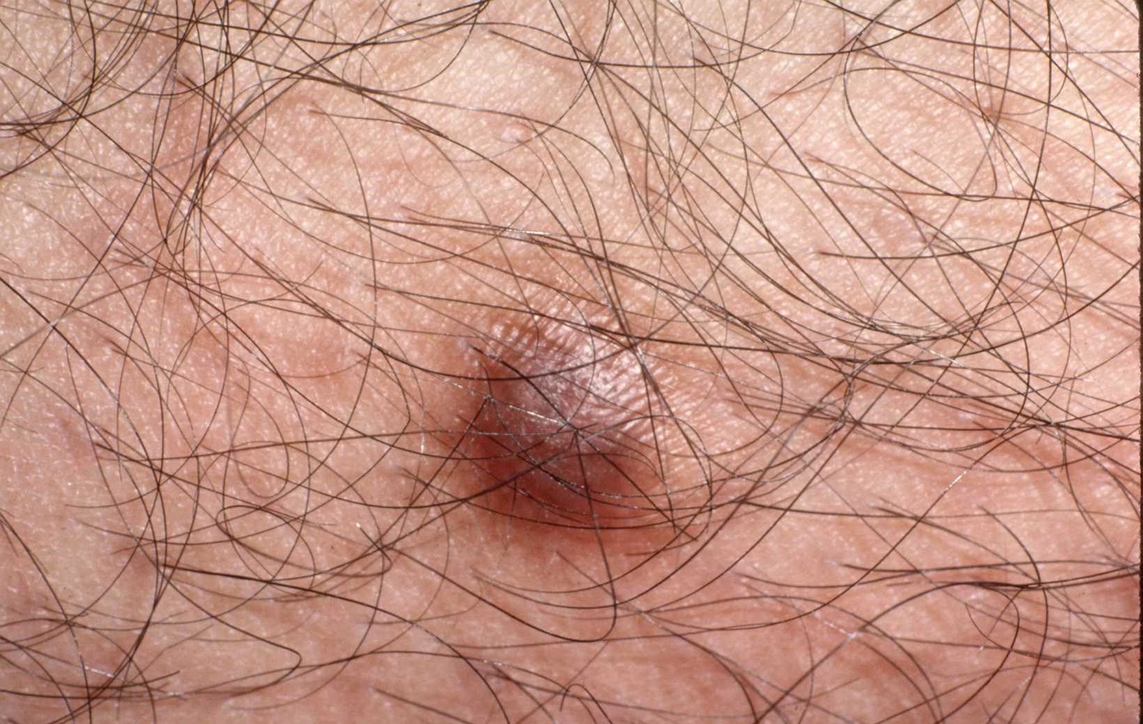
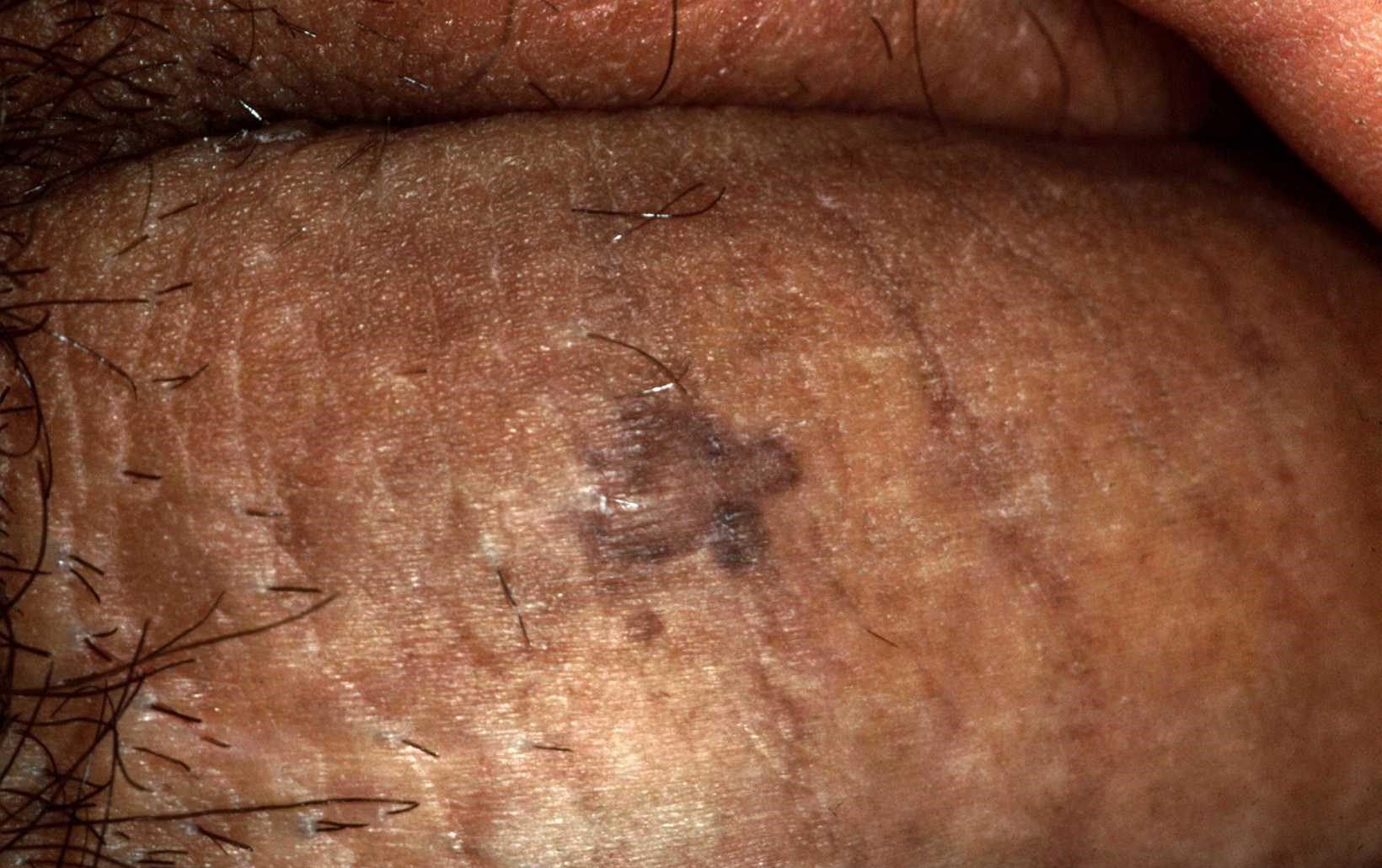
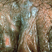
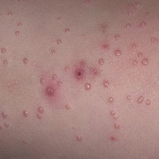
Bowenoid papulosis is now most commonly known to occur on the genitalia of both sexes in sexually active people. Bowenoid papulosis is manifested as papules that are induced virally by human papillomavirus (HPV) and demonstrate a distinctive histopathology (bowenoid dysplasia). Many bowenoid papulosis lesions appear to run a benign course, although a number of case reports associate bowenoid papulosis with malignant invasive transformation (2.6%).
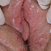
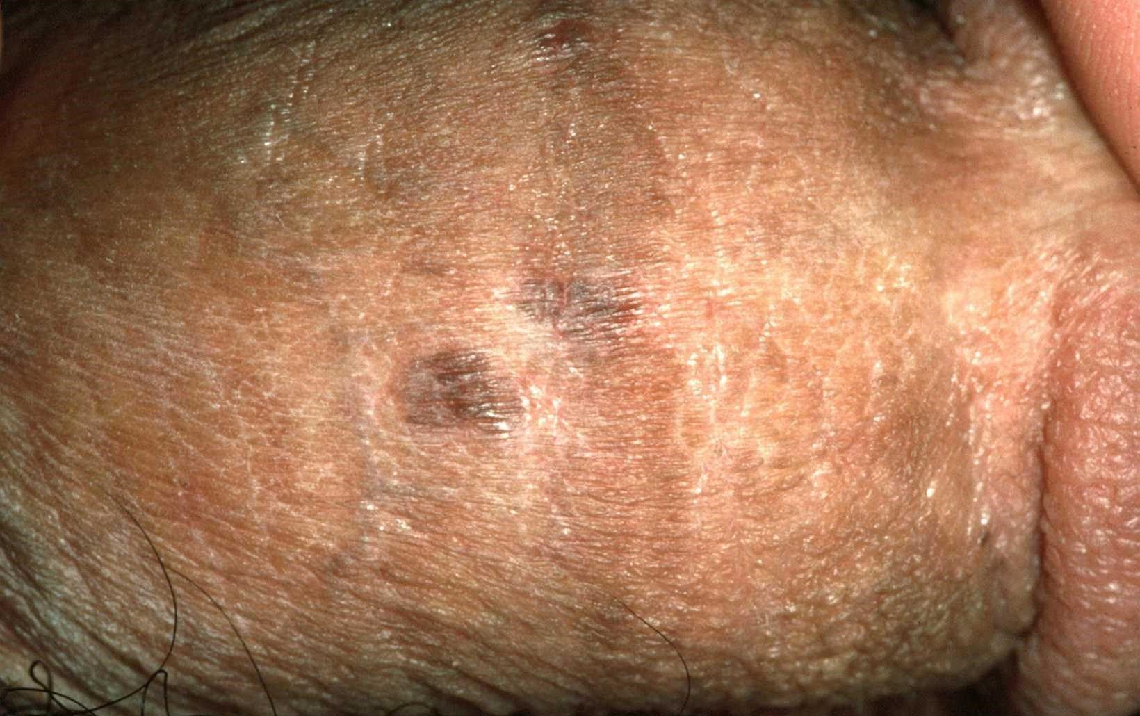
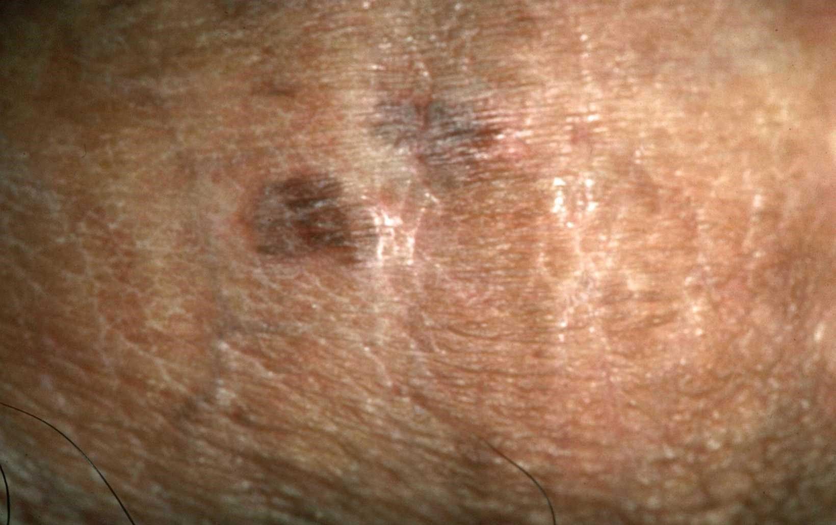
Bowenoid papulosis may be considered to be a transitional state between a genital wart and Bowen disease. The rate of transformation of bowenoid papulosis lesions is unknown. Clearly, bowenoid papulosis lesions have some malignant potential, but they may be treated with locally destructive modalities, sparing the surrounding tissues. Bowenoid papulosis lesions often are multifocal, and patients should be observed for recurrence and for the possibility of invasive or in situ malignancy.
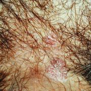
Bowenoid papulosis is an asymptomatic focal epidermal hyperplasia and dysplasia induced by HPV infection (most commonly by HPV 16). The result can appear as a papule or multiple papules that sometimes coalesce, as patches, or as plaques. Histologically, they are composed of scattered atypical cells or full-thickness epidermal atypia that some view as analogous to squamous cell carcinoma in situ. This epidermal atypia is sometimes known as bowenoid dysplasia.
Bowenoid papulosis occurs primarily in young, sexually active adults, with a mean age of 31 years. Because Bowenoid papulosis usually runs a benign course with many cases spontaneously regressing, treatment is often unnecessary. Lesions should be re-examined every 3 to 6 months so that any changes may be picked up early.